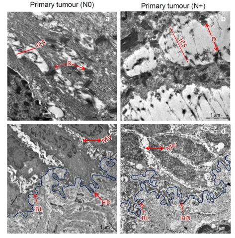
Cell adhesion is of crucial importance in the maintenance of tissue integrity and hence any alterations in cell adhesion molecules and/or ultrastructure have potential to influence the processes of neoplastic transformation as well as progression. Although, it is suggested that disruption of cellular adhesion is a hallmark of malignant transformation, biological evidence is not available. Owing to the high resolution, transmission electron microscope can detect subcellular changes at early stage of disease. Study is designed to evaluate the potential of electron microscope (EM) in early assessment of oral leukoplakia lesions for malignant transformation and progression using quantitative EM technology-iTEM software. Ultrastructural alterations were detected in the desmosomes, hemidesmosomes and basal lamina at hyperplastic grade and their severity significantly increased as the disease progressed. Some of the leukoplakia/tumours diagnosed as lower grade using histopathology, appeared to be higher grade when observed under EM. This was confirmed by clinical follow-up in OSCC patients.
This study highlights the role of EM in detecting subcellular morphological changes which could not be detected on histopathological examination at an early stage of disease. Reduction in desmosome, hemidesmosome number and increased length of basal lamina could be the criteria for the assessment of malignant potential of leukoplakia lesions.


