Inner Banner

Arvind Ingle group
Dr. Arvind Ingle

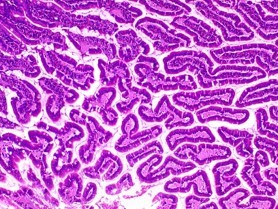
The Histology facility provides the following services to the Centre: (a) slides of unstained/ haematoxylin and eosin (H&E) stained histology sections of animal tissues including bone/ tumour samples, (b) logistic support for frozen sectioning of human/ animal tissues, and (c) blocks of multiple tissues by pecking method using a microarray machine. During 2017, the facility received 3471 tissue samples in fixative and 1231 human tissue paraffin blocks and, after processing, supplied 6869 stained and 17681 unstained slides to 20 research labs. In addition, 3397 tissues were processed for cryo-sectioning, and 1318 H&E stained and 1318 unstained slides were supplied to 12 research labs.

Dr. Sanjeev Waghmare
Facility In Charge: Ms. Shamal Vetale
About Us

The Flow Cytometry (FCM) facility is a centralized facility used by scientists/ clinicians from ACTREC for a wide range of research applications including Immunophenotyping, Multicolor analysis, DNA content and cell cycle analysis, Apoptosis and Proliferation studies, Detection of mitochondrial membrane potential, Stem cell analysis, Detection of circulating tumor cells, Functional assays like Intracellular calcium influx, Oxidative burst analysis, Intracellular cytokine analysis, Cytometric bead array assay for the detection of cytokines, and 4-way live cell sorting.

The facility provides technical expertise in experiment design and data interpretation to researchers whenever required, and also provides training in data analysis.

The facility has four flow cytometers: FACSAria III, FACSAria I, Attune NxT and FACSCalibur. FACSAria III is equipped with 5 lasers (UV: 355 nm, Violet: 405 nm, Blue: 488 nm, Yellow green: 561 nm and Red: 633 nm) and can perform 18 color analysis. FACSAria-I is equipped with 3 lasers (Violet: 405 nm, Blue: 488 nm and Red: 633 nm) and can analyse up to 11-color. Both the Cell sorter can perform 4-way cell sorting as well as single cell sorting into 96-well plates using the Automated Cell Deposition System. Attune NxT is equipped with 4 lasers (Violet: 405 nm, Blue: 488 nm, Yellow green: 561 nm and Red: 633 nm) and can analyse up to14 color. FACSCalibur is equipped with one laser (Blue: 488 nm) and can perform 3-color analysis.

The software used for data analysis include FlowJo, FACSDiva, CellQuest Pro, Attune NxT, FCAP Array and Modfit.

The facility also offers its services to outside investigators on payment basis. Demonstrations and training were provided to staff and students on request.

Services

Flow cytometry facility is a centralised facility provides technical resources and professional expertise for multicolour flow cytometry analysis and cell sorting.

The following services has been provided by Facility:

  • Multicolor analysis can be done efficiently up to 18 colors by FACSAria III (BD Biosciences) and 14 colors by using Attune NxT (Life Technologies).
  • Cell sorting can be done by FACSAria I (up to 11 Colors and FACSAria III (up to 18 Colors). Both the cell sorters can sort 4 different populations at a time and also perform plate sorting for clonal analysis.
  • Software Flow Jo, FACSDiva, ModFit (Cell cycle analysis), Attune NxT, Cell Quest Pro, FCAP Array (Cytometric Bead Array Assay analysis) can be used for analysis purpose.
  • The technical support is extended to design the flow cytometry based experiments, to optimise the reagents (e.g. antibodies concentration), Florochrome selection and further assistance in data analysis.

For External User

The Flow cytometry facility at ACTREC is open to external user with certain conditions as follows:

  • Booking has to be done at least one week prior in advance and depending upon the availability of the slot, booking will be available to external user.
  • User should prepare the sample in their own Institute/ Lab and bring it in sterile condition.
  • It is recommended to pass the sample through strainer to avoid clogging of sample line.
  • The user should bear the cost of analysis of data separately.
  • The data should be collected immediately after acquisition. Users are requested to carry CD to copy the data.

Charge for Outside Users

Government Institutes/ University   - Rs. 600/ Tube (for acquisition)
Corporate                  - Rs. 2000/ Tube (for acquisition)

The user should contact of any query to Flow Cytometry facility on following:

Dr. Sanjeev Waghmare
Email: swaghmare@actrec.gov.in
      flowcytometry@actrec.gov.in
Contact: 022-27405000 Ext. 5316/5122

Protocol

DNA Cell Cycle using Propidium iodide

Reagents and Instruments

  • Cell lines or primary cell cultures
  • Phosphate buffered saline (0.01 M PBS pH 7.4)
  • 70% ethanol
  • Propidium iodide (PI)- Stock 400 μg/ml
  • RNase- Stock 1mg/ml
  • Triton X-100
  • Polystyrene round bottom 12 x 75 mm Falcon tubes (Flow Tubes)
  • 1 ml syringes with 26 G (Guage) needle
  • Flow cytometer- FACSCalibur, Attune Nxt
  • Softwares – CellQuest Pro, ModFit

Sample fixation

  1. Dispense 1 x 10⁶ cells per ml of PBS in 5ml polystyrene tubes.
  2. Wash the cells with 1 ml of PBS by centrifuging at 1000 rpm for 10 mins.
  3. Fix the cells by adding 70% chilled ethanol drop wise to the cell pellet while vortexing. This should ensure fixation of all cells and minimize clumping.
  4. Keep cells on ice for 1 hr or overnight at 4℃. (Fixed cells can be stored at 4℃ for 15-20 days)

Sample Staining

Procedure – I
  1. Take 1 x 10⁶ fixed cells in 5ml polystyrene tubes and centrifuge at 1000 rpm for 10 mins.
  2. Discard supernatant, wash the cells with 1 ml of PBS by centrifuging at 1000 rpm for 10 mins.
  3. Add 500 μl of PBS to the cell pellet, vortex gently, add 100 μl of Propidium Iodide and 100 μl of RNase. Cover the tubes with aluminum foil to prevent its exposure to light. Incubate at 37ºC for 30 mins.
  4. Pass the sample through 26 G needle to prepare single cell suspension.
  5. Acquire the sample on FACSCalibur / Attune NxT.
Procedure- II
  1. Take 1 x 10⁶ fixed cells in 5ml polystyrene tubes and centrifuge at 1000 rpm for 10 mins.
  2. Discard the supernatant and wash the cells with 1 ml of PBS by centrifuging at 1000 rpm for 10 mins
  3. Re-suspend the pellet in 500 μl of PBS containing 0.1% Triton X-100 and 100 μg/ml of RNaseA for permeabilization and RNase inactivation respectively. Incubate at 37ºC for 15 mins.
  4. After incubation, add 25 μg/ml of propidium iodide to the cell suspension, mix properly and further incubate at 37°C for 15 minutes.
  5. Pass the cell suspension through 26 G needle to avoid clumping of sample.
  6. Acquire the sample on FACSCalibur / Attune NxT.

Preparation of reagents

1. Phosphate buffer saline (0.01 M PBS pH 7.4)

NaCl8.0 g
KCl0.2 g
Na2HPO41.44 g
KH2PO40.24 g

Dissolve in 800 ml distilled water (dH2O)
Adjust the pH to 7.4 with HCl
Finally, make up the volume to 1 L with dH2O.
Store at 4°C.

2. 70 % ethanol

Ethanol70 ml
Distilled water (dH2O)30 ml

Store at 4°C.

3. Propidium iodide (PI): Stock – 400 μg /ml

Propidium iodide (Sigma)4 mg
PBS (0.01 M, pH 7.4)10 ml

Dissolve and Aliquots in 1.5 ml Eppendorf tubes
Store at -20°C.
(Precaution: Cover the 1.5 ml Eppendorf tubes with Aluminum foil as PI is light sensitive.

4. RNase: Stock – 1 mg/ml

RNase (Sigma)1 mg
PBS (0.01 M, pH 7.4)1 ml

Dissolve and aliquot in Eppendorf tubes.
Store at -20°C.

Point to remember:

  1. The quality of sample should be maintained for better result.
  2. Try to make single cell suspension before fixation of the sample to avoid clumping (to prevent the doublets).
  3. Handle the tubes with care and wear gloves to avoid contact with skin as PI is mutagenic.

Immunophenotyping

There are three general staining protocols based on type of antigen to be evaluate, nature of Fluorochrome and sample type.

  1. Direct Staining:

    This technique involves incubation of cells with fluorochrome conjugated antibody, which recognize and bind to various cell surface antigens. The antibodies are mostly monoclonal antibodies (MAb) and they may be added either single or in multiple combinations depending upon the analysis.

  2. Indirect staining:

    In indirect staining technique, the cells are incubated with an unconjugated primary antibody. The primary antibody recognizes and binds to cell surface antigens then the cells are subsequently incubated with a fluorochrome conjugated secondary antibody.

  3. Intracellular staining:

    This technique involves detection of intracellular antigens; it done by direct staining method using conjugated antibodies. Cells are initially subjected to a fixation step with paraformaldehyde in order to minimize leakage of proteins out of the cell, following permeabilization by mild detergents such as saponin or Tween 20. The permeabilization allows access of conjugated antibodies to proteins within the cell. The conjugated antibody penetrates and binds to its target within the cell. Intracellular staining technique is very useful in monitoring immune responses and tracking signalling molecules.

Reagents and Instruments

  • Cell line or primary cells
  • Phosphate buffered saline (PBS, 0.01M pH 7.4)
  • FACS Buffer I (for surface staining)
  • FACS Buffer II (for intracellular staining)
  • 1% Paraformaldehyde
  • Refrigerated centrifuge
  • Polystyrene round bottom 12 x 75 mm Falcon tubes
  • Micropipettes of capacity- 1 ml, 200 μl, 10 μl
  • Flow cytometer – FACSCalibur, Attune NxT or FACSAria
  • Software - CellQuest Pro (FACSCalibur), Attune NxT (Attune NxT), FACSDiva (FACSAria)
Antibodies

Direct staining:
1. Fluorochrome labelled antibody
2. Isotype matched fluorochrome labelled antibody
Indirect staining:
1. Unlabelled primary antibody
2. Fluorochrome labelled secondary antibody

Procedure

  1. Direct Staining
    1. Dispense 1 x 10⁶ Cells in 5ml polystyrene tubes.
    2. Add 1 ml of FACS buffer I and centrifuge the tubes at 1000 rpm for 10 mins at 4°C. Discard the supernatant. (washing)
    3. Resuspend the cells in 100 μl of FACS buffer I.
    4. Add appropriate amount of antibody (the amount of antibody can be decided as per manufacturer’s recommendation and titration)
    5. As negative control, incubate the cells with isotype matched antibody conjugated with same fluorochrome used for the test.
    6. Incubate the cells on ice at 4°C for 45 mins. in dark.
    7. Wash the cells 2 times and finally resuspend in 500 μl of FACS buffer I.
    8. Acquire the sample on flow cytometer as soon as possible.
    9. If the acquisition is not possible on the same day, fix the cells in 500 μl of 1% paraformaldehyde to preserve the cells for several days (this will stabilize the light scatter, prevent quenching and inactivate most biohazardous agents).
  2. Indirect Staining
    1. Dispense 1 x 10⁶ Cells in 5ml polystyrene tubes.
    2. Add 1 ml of FACS buffer I and centrifuge the tubes at 1000 rpm for 10 min at 4°C. Discard the supernatant (washing).
    3. Resuspend the cells in 100 μl of FACS buffer I.
    4. Add appropriate amount of unconjugated purified monoclonal antibody specific for antigen to be detected. (the amount of antibody can be change based on manufacturer’s recommendation and titration)
    5. Incubate on ice at 4 0 C for 45 mins.
    6. Wash the cells 2 times with FACS buffer I as described before to remove excess antibody.
    7. Dilute the fluorochrome- labelled secondary antibody as per manufacturer’s instruction in FACS buffer I.
    8. Incubate the cells with diluted secondary antibody (specific for primary antibodies) conjugated with fluorochrome on ice at 40 C for 45 min.
    9. As negative controls, incubate the cells with isotype matched controls conjugated with same fluorochrome used for the test.
    10. Wash the cells 2 times and finally resuspend in 500 μl of FACS buffer I.
    11. Acquire the sample on flow cytometer as soon as possible.
    12. If the acquisition is not possible on the same day, fix the cells in 500 μl of 1% paraformaldehyde to preserve the cells for several days (this will stabilize the light scatter, prevent quenching and inactivate most biohazardous agents).
  3. Intracellular staining
    1. Dispense cells in 5ml tubes.
    2. Add 1 ml of FACS buffer I and centrifuge the tubes at 1000 rpm for 10 mins at 40C. Discard the supernatant (washing).
    3. Add 500 μl of 1 % paraformaldehyde to fix the cells.
    4. Incubate on ice at 40C for 15 mins. with intermediate mixing of the tubes.
    5. Wash the cells with FACS buffer I by centrifuging tubes at 1000 rpm for 10 mins. at 40C twice.
    6. After second wash, Add 100 μl FACS buffer II to the cell pellet to permeabilize the cells.
    7. Gently vortex the tubes.
    8. Incubate at room temperature for 5 min.
    9. Add recommended amount of fluorochrome labelled antibody specific for antigen to be detected.
    10. As negative control, incubate the cells with isotype matched controls conjugated with same fluorochrome used for the test.
    11. Incubate on ice at 4°C for 45 mins. in dark.
    12. Wash the cells 2 times and resuspend in 500 μl of FACS buffer I.
    13. Acquire the sample on flow cytometer.

Preparation of reagents:

1. Phosphate buffer saline (0.01 M PBS pH 7.4)

NaCl8.0 g
KCl0.2 g
Na2HPO41.44 g
KH2PO40.24 g

Dissolve in 800 ml distilled water (dH2O)
Adjust the pH to 7.4 with HCl
Finally, make up the volume to 1 L with dH2O.
Store at 4°C.

2. FACS buffer I

PBS (0.01 M, pH 7.4)99 ml
Fetal Bovine Serum (Invitrogen)1.0 ml
Sodium Azide (NaN3)0.02 g

Store at 4°C.

3. FACS buffer II

PBS (0.01 M, pH 7.4)99 ml
Fetal Bovine Serum1.0 ml
Sodium Azide (NaN3)0.02 g

Store at 4°C.

4. 1 % paraformaldehyde

Paraformaldehyde1.0 g
PBS (0.01 M, pH 7.4)100 ml

Prepare solution in a glass bottle. Heat in microwave oven until the powder dissolves completely. Allow it to cool. Cover the bottle with Aluminium foil to prevent exposure to light. Store at 4°C.
(Precaution: Do not inhale the fumes)

Point to remember:

  1. Before starting the experiment, the titration of antibodies should be done according to manufacturer’s instruction for optimum concentration.
  2. The recommended controls should be run with every experiment for better analysis e.g. Iso control
  3. In case of multicolour analysis use compatible compensation controls.

Annexin V / Propidium iodide staining (For Apoptosis)

Reagents and Instruments:

  • Phosphate buffered saline (0.01 M PBS pH 7.4)
  • Annexin-V FITC
  • Propidium iodide (PI) – stock- 400 μg/ml
  • Annexin Binding buffer- 10X
  • Refrigerated centrifuge
  • Polystyrene round bottom 12 x 75 mm Falcon tubes
  • Micropipettes of capacity- 1 ml, 200 μl, 10 μl
  • Flow cytometer- FACSCalibur, Attune Nxt
  • Softwares – CellQuest Pro, Attune NxT, Flow Jo

Procedure:

  1. Wash the cells twice with 1X PBS and then resuspend in 1X Annexin Binding Buffer at a concentration of 1 X 10⁶ cells/ml.
  2. Transfer 100 μl of the cell suspension (1 X 10⁵ cells) in another polystyrene tube.
  3. Add 2-5 μl of Annexin V FITC.
  4. Add 40-50 μg of Propidium Iodide.
  5. Gently vortex the cells and incubate for 15 min at room temperature in the dark.
  6. Add 300 μl of 1X Annexin Binding Buffer to each tube. Analyze by flow cytometry within 1 hr.
The following controls are used to set up compensation
  1. Unstained cells.
  2. Cells stained with only Annexin V FITC
  3. Cells stained with only Propidium Iodide

Preparation of Reagents:

1. Phosphate buffer saline (0.01 M PBS pH 7.4)

NaCl8.0 g
KCl0.2 g
Na2HPO41.44 g
KH2PO40.24 g

Dissolve the following in 80ml MilliQ Water:

2. Annexin binding buffer 10X

HEPES (0.1 M)2.603 g
KCl0.2 g
cacl20.368 g
NaCl (1.4 M)8.182 g

Adjust the pH to 7.4. Make up the volume to 100 ml with MilliQ water. Store at 2-8°C

3. Propidium iodide (PI): Stock – 400 μμg /ml

Propidium iodide (Sigma)4 mg
PBS (0.01 M, pH 7.4)10 ml

Dissolve and aliquot in 1.5 ml capacity tubes.
Store at -20°C.
(Precaution: Cover the tube with aluminium foil as PI is photo-sensitive.)

Point to remember:

  1. Before starting the experiment, the titration of Annexin V should be done to decide optimal concentration.
  2. The pH of Annexin binding buffer should be maintained for better results.
  3. Handle with tubes with care and wear gloves to avoid contact with skin as PI is mutagenic.
User Guidelines

SOP for Flow Cytometry Facility User

New Flow Cytometry facility users are required to fill up the Project details form before starting the experiment. A hard copy of the same should be submitted with signatures of Principal Investigator/Officer-in-charge/ Scientific officers. The format will be available at the Facility.

Booking for Acquisition and Analysis of samples

  1. Booking has to be done in Booking register that is available in the Facility.
  2. Booking can be done one-week prior to the date of sample acquisition.
  3. Booking slots are available for both acquisition and sorting. In a given day, only one booking slot of 2 hrs is allowed. Additional time will be entertained based on the nature of the experiments and number samples depending on the availability of the slots.
  4. Cancellation of booking if any should be informed at least one-day prior of date of acquisition.
  5. If any queries, please contact Facility staff.

Sample Preparation for Flow Cytometry

  1. Try to prepare single cells suspension and avoid clumping. Users are Recommended to pass the cells through filters or strainers or insulin syringe.
  2. Sample concentration should be maintained for better results. (e.g. 1 X10⁶ Cells / 300-500 μl for acquisition depending upon the cell type)
  3. For sorting carry sufficient number of collection tubes with appropriate quantity of media or buffer.
  4. User can use 15 ml tubes, 5 ml FALCON tubes, Eppendorf 1.5 /2 ml, and 96 well plates for collection of cells.

General Guidelines

  • Suitable Controls should be carry at the time of acquisition and if any queries please contact Facility Staff.
  • Data can be collected in FCS format in the CD immediately after the sample acquisition.
  • Users can use softwares Flowjo, FACS DIVA, Attune NxT, Modfit LT 5.0, Cells Quest Pro, for analysis with prior booking.
Contact Us

Dr. Sanjeev K Waghmare
Principal Investigator
Scientific Officer’ F’
Officer In-Charge
Email: swaghmare[at]actrec[dot]gov[dot]in
Phone:022-27405122

Flow Cytometry Facility, ACTREC
Email: flowcytometry[at]actrec[dot]gov[dot]in
Phone: 022-27405000 Ext. 5316


EM gp
Facility In Charge: Siddhi A. Redkar

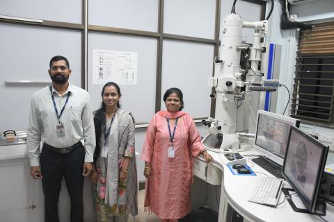
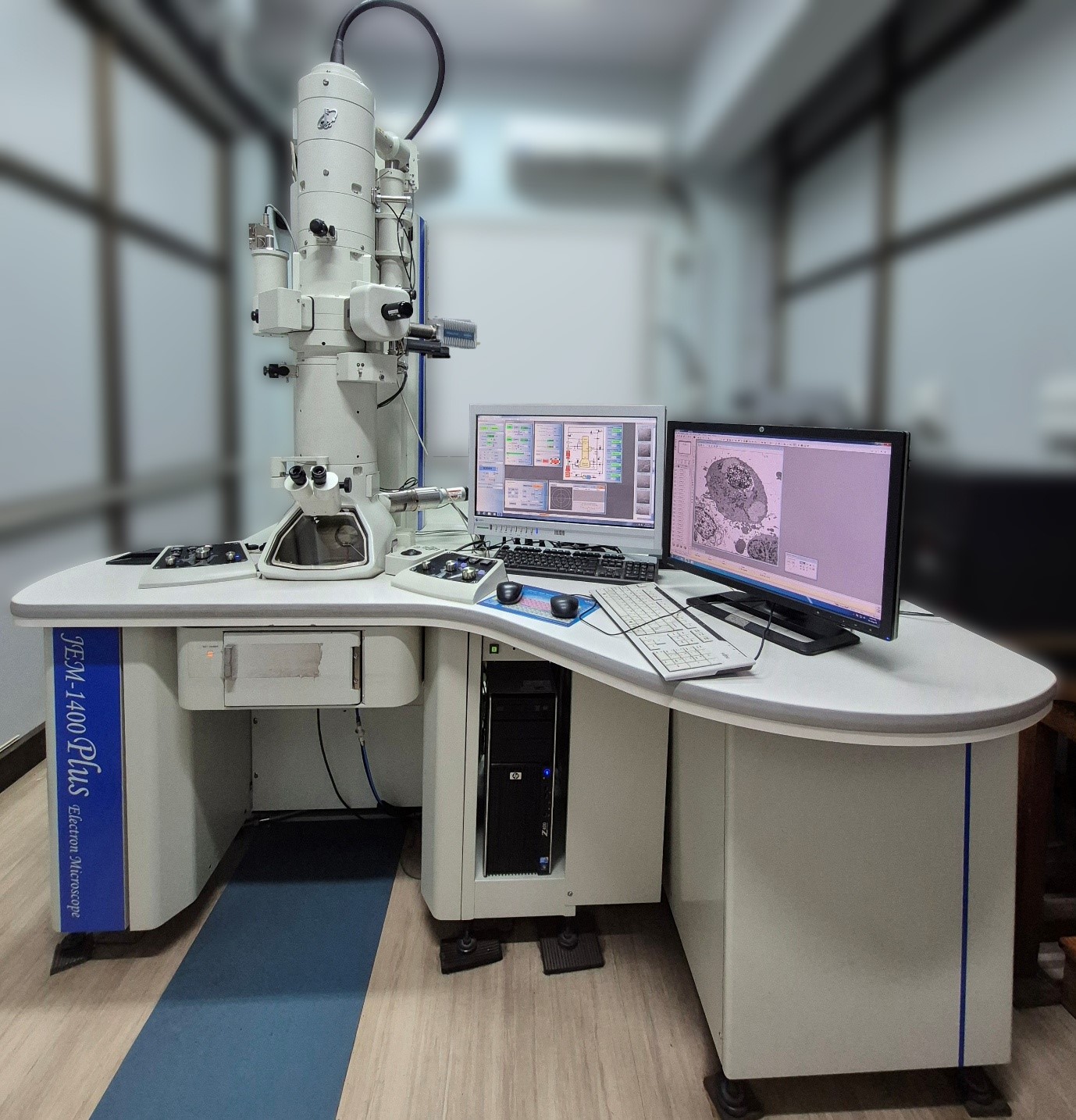
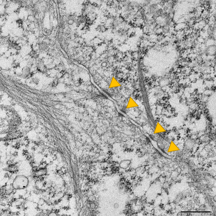
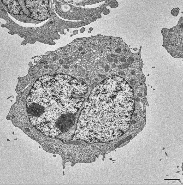
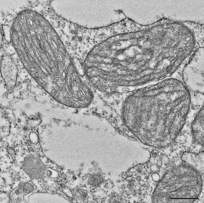
The Electron Microscope Facility at ACTREC is equipped with JEM 1400 PLUS, a Transmission electron microscope (JEOL, Japan) functioning at 120kV with magnification up to 12,00,000 X and 0.2nm resolution. TEM analysis provides a detail ultra- structural information of resin embedded or negatively stained biological samples. The facility extends its services for research and analysis of biological samples, nanoparticles and other applications to the external researchers along with the in-house researchers. The facility is also equipped with instruments such as ultra-microtome, glass knife-maker, auto-trimmer and glow discharge unit.

Equipments

TRANSMISSION ELECTRON MICROSCOPE Make: JEOL Model: JEM 1400 PLUS

equipped


JEM 1400 PLUS is a Transmission electron microscope (JEOL, Japan) functioning at 120kV with magnification up to 12,00,000 X and 0.2nm resolution.

Specifications

Accelerating voltage 120 kV
Electron SourceTungsten filament
ResolutionHC pole piece
Point image0.38 nm
Lattice image0.2 nm
Magnificationx200 to x1200000
CameraBottom Mounted Olympus TENGRA 5.3 MP CCD Camera
Tilt range±70°

Applications

  • Imaging of resin embedded cells and tissues.
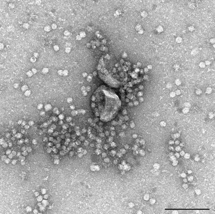
  • Imaging of negatively stained small particles (<100nm) like bacteriophages, exosomes, nanoparticles, proteins and DNA.
  • Acquisition of immunogold labelled macromolecules to detect specific localization.
  • Elemental detection by Bruker EDS system.

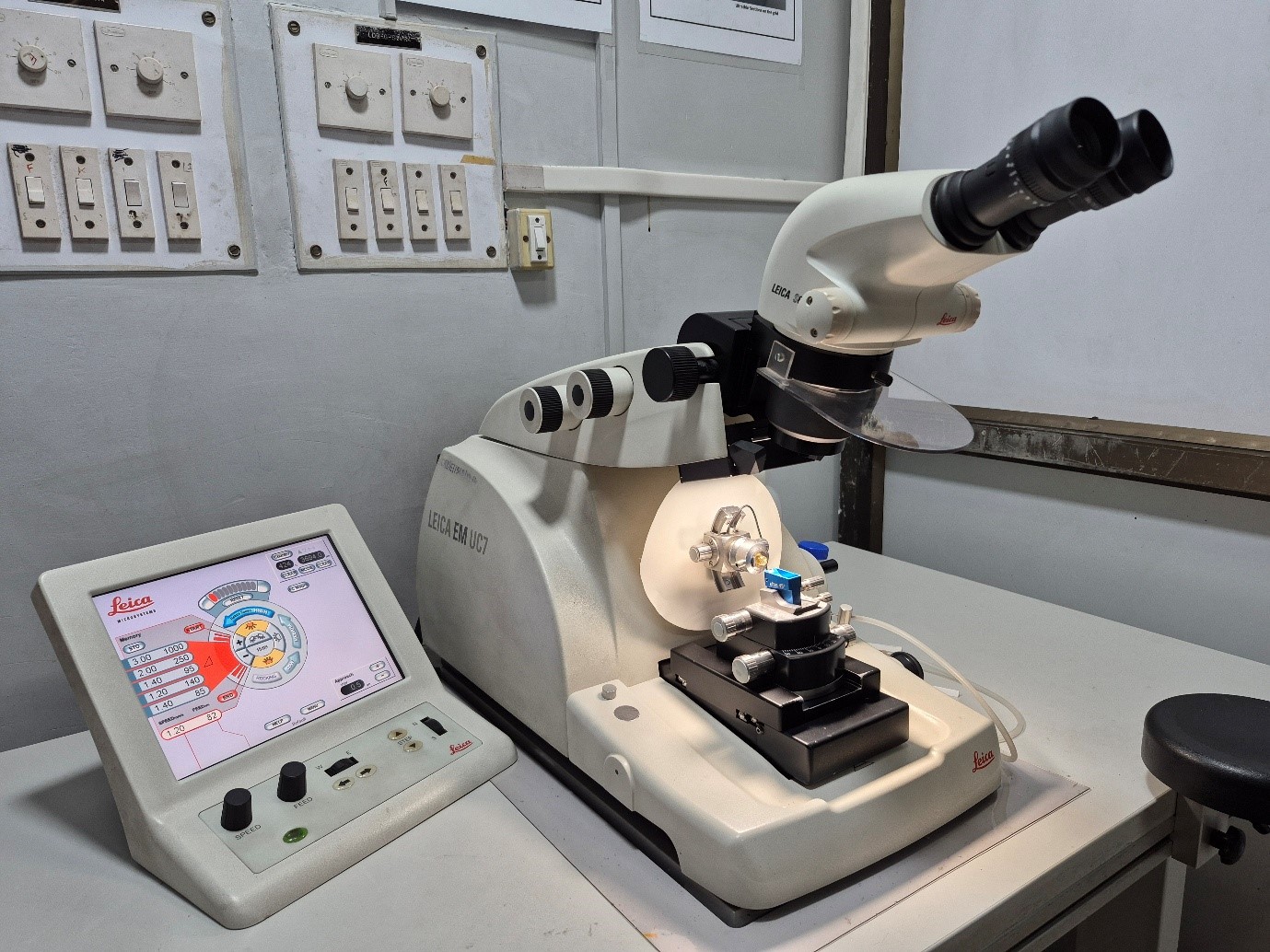
EQUIPMENT: ULTRAMICROTOME Make: Leica Model: UC7

equipped

Ultramicrotome provides high-quality preparation of ultra-thin or semi-thin sections for transmission electron microscopy. The desired thickness of the section can be selected with touchscreen control unit and sections can be cut using glass or diamond knives with motorized knife stage. Majorly, 70nm ultra-thin sections are cut for electron microscopy.

Application

  • Ultra-thin and semi-thin sectioning of resin embedded cells and tissues.

Additional equipment’s

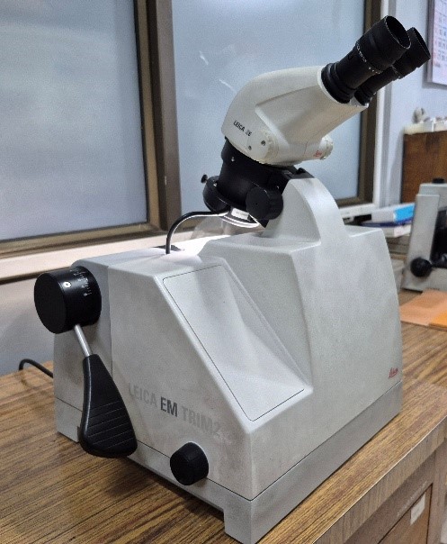
equippedLeica EM TRIM2 auto-trimmer – for trimming the resin blocks,
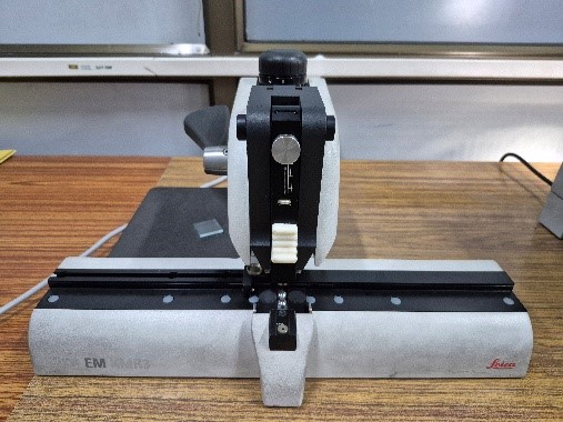
equippedLeica EM KMR3 – Glass knife maker – for making glass knives.
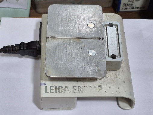
equippedLeica EM MP Hotplate – for heating the resin sections.

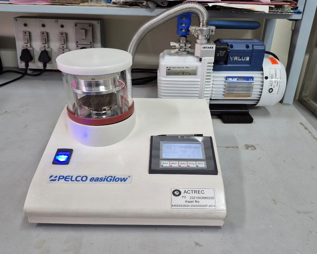
GLOW DISCHARGE UNIT Make: Ted Pella Model: PELCO easiGlow

equipped


The PELCO easiGlow uses an automated and quick cycle with fully selectable parameters for one of the most common glow discharge applications, making TEM support films or grids hydrophilic using air.

Applications

  • To make TEM support films or grids hydrophilic using air
  • Cleaning TEM grids
  • Adhesion and orientation of proteins, nucleic acids and extra cellular vesicles.
  • TEM grid preparation for nanoparticle studies
Current Applications
  • The facility carries out sample preparation for routine electron microscopy, including resin block making (solid tissues, monolayer cell cultures, and cell suspensions) followed by ultrathin sectioning using Leica UC-7 ultramicrotome, contrasting, and acquisition.
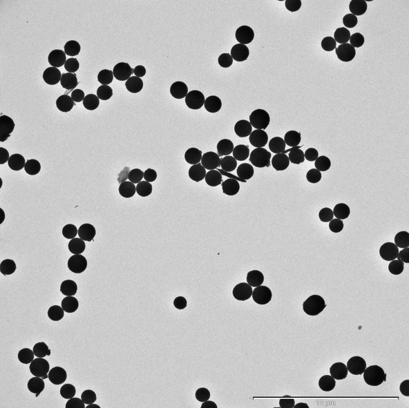
  • The facility also carries out negative staining for small particles (<100nm) like bacteriophages, exosomes, nanoparticles, proteins and DNA.
  • The facility provides acquisition of immunogold labelled macromolecules to detect specific localization.
  • The facility provides elemental detection by Bruker EDS system

The major areas of electron microscopy analysis are,

  • Ultrastructure study of cell and tissue,
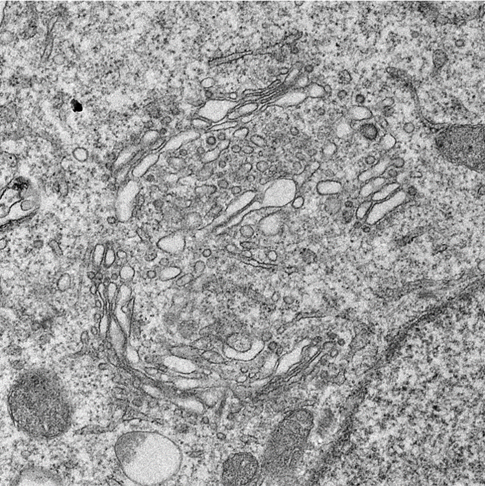
  • Studying the details at organelle level like mitochondrial changes, ER - mitochondrial interactions, mitochondrial cristae, cell membrane, nuclear architecture, golgi complexes etc.,
  • To check events of autophagy, apoptosis or necroptosis,
  • Characterization of exosomes, bacteriophages and nanoparticles,
  • Uptake and effect of nanoparticles / drugs / radiation on cells or tissue,
  • EDS analysis of nanoparticles
Electron Microscopy ATLAS
Sample Submission

External users: Please write to the facility regarding your requirement for electron microscopy. The facility will provide the details as per the analysis requirement. The users are requested to discuss over email regarding their study and sample submission followed by booking a slot accordingly. The samples need to be submitted along with a duly filled requisition form. For quotation, a duly filled requisition form can be mailed to the facility email address (tem.facility@actrec.gov.in). For requisition form, please click the link below,

Internal users (ACTREC):

  • For routine EM, please check with the facility regarding the sample submission requirements as per the study.
  • For negative staining, please book a slot after discussing with the facility for sample submission. Only three samples will be accepted per slot.
  • Samples should be accompanied by a duly filled requisition form and online requisition.

    Requisition form

Charges
ACTREC & DAEOther than ACTREC institutionsCorporate
Routine EM1000500010000
Routine EM + Immunogold labeling1500600012000
Negative staining80040008000

18% GST to be included in addition to the following charges.

Mode of Payment

External users: The users should pay the charges against the quotation or invoice to the following account within a week of the invoice. The users should share the transaction details with the facility once the payment is done. The acquisition will be carried out after the receipt of the payment is confirmed. For e-payment details please click the link below.

Epayment details

Contact

Facility-in-charge:
Siddhi A. Redkar
Technical Officer ‘D’
Electron Microscope Facility
(Khanolkar Shodhika - 68)
ACTREC, TMC
Plot No.1 & 2, Sector 22
Kharghar, Navi Mumbai - 410 210.
022-27405000 / 022-68735000
Extension : 5545
Email : tem.facility@actrec.gov.in

EM Staff:
Ms. Arpana Kadam, Scientific Assistant ‘C’
Mr. Shridhar Nadkar, Technician ‘F’

Media Gallery
CellCell
Cell to cell ContactCell
CellCell
ExosomesMitochondria
CellCell
NanoparticlesGolgi Complex
Publications

2026


Dr. Pradnya Kowtal
Officer In Charge: Dr. Pradnya Kowtal

The DNA sequencing facility has two automated DNA sequencers: an eight capillary Genetic Analyzer 3500 and a 48 capillary Genetic Analyzer 3730 from Applied Biosystems/ Thermofisher, both of which are used for DNA sequencing and fragment analysis. The average turnaround time to give out data is one working day after receiving samples. During the year 2017, the facility carried out a total of 25,459 reactions of which 13,718 were for sequencing and 11,741 were for fragment analysis. The facility also provided demonstration of Sanger sequencing to students and other visitors.

Digital Imaging
Facility In Charge: Mrs. Vaishalii kailaje

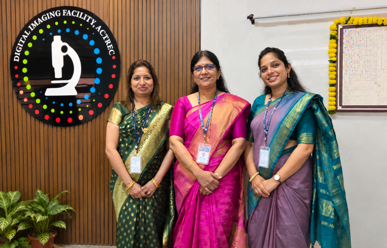
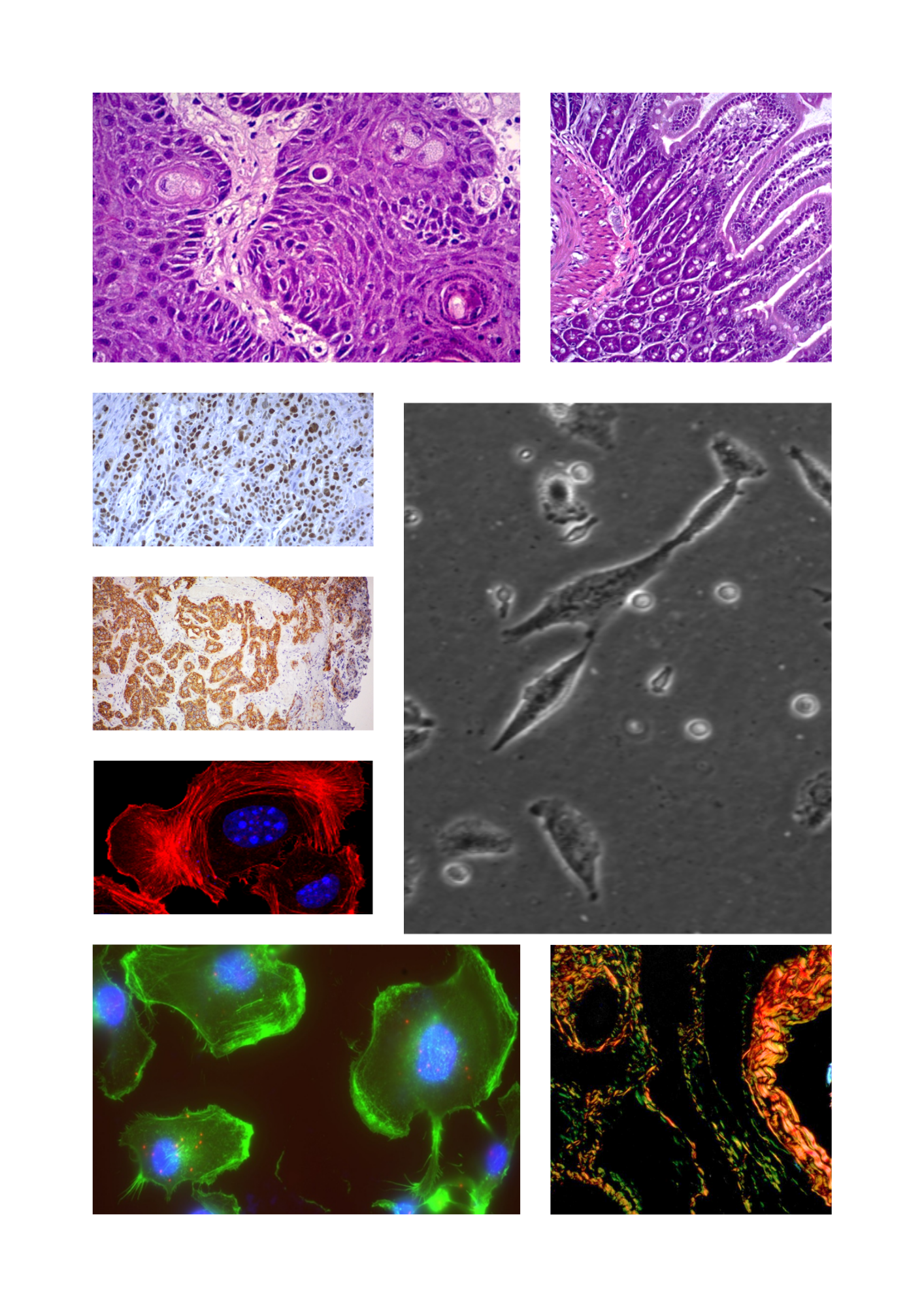
The Digital Imaging Facility is a centralized core facility that provides advanced microscopy and image analysis services to students and researchers across the institute. By integrating state-of-the-art imaging technologies with powerful image analysis tools, the facility enables researchers to visualize, quantify, and gain deeper insights into complex biological structures and processes.

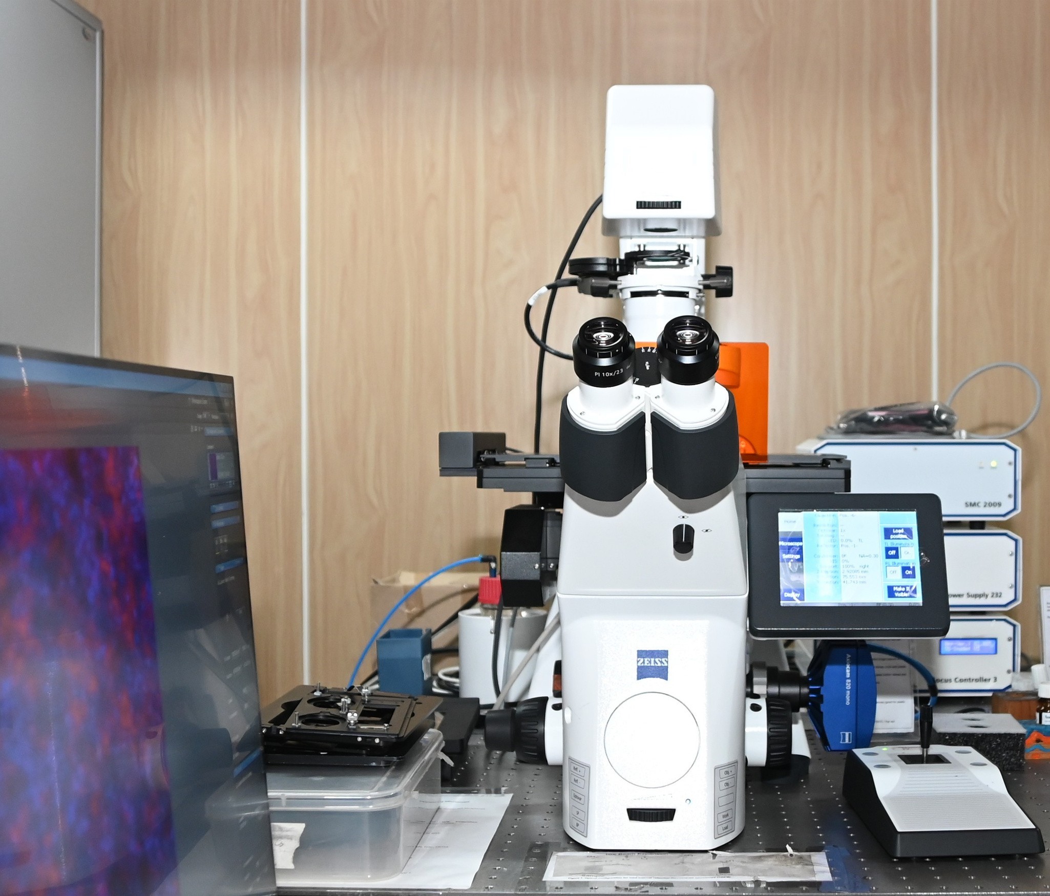
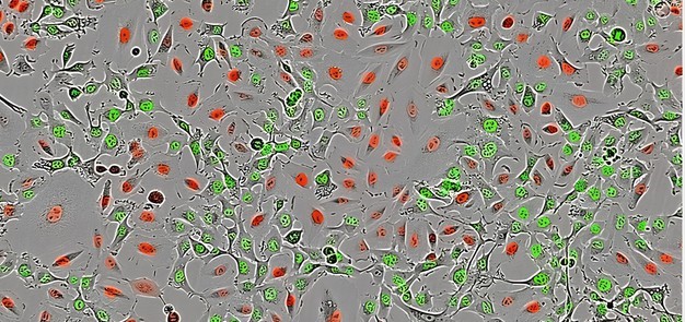
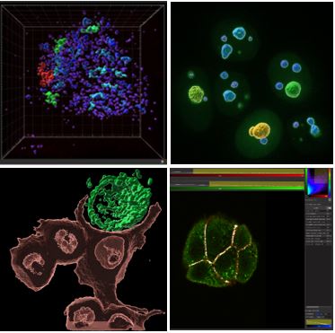
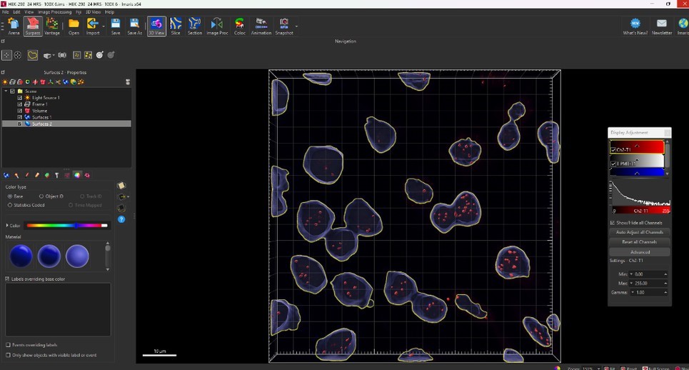
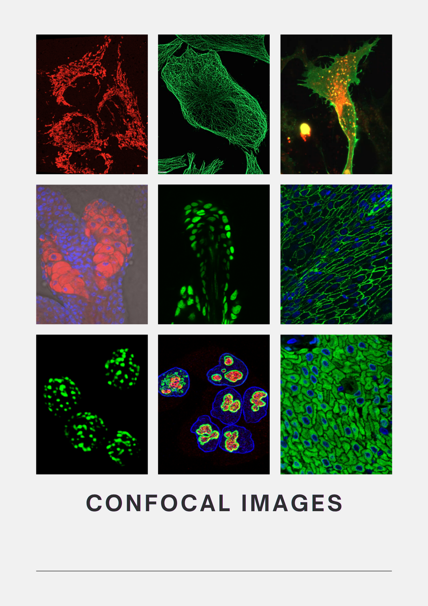
The facility offers a comprehensive range of imaging platforms, including five widefield microscopes, three confocal microscopes all with dedicated live-cell imaging capabilities, and the Incucyte Live-Cell Imaging and Analysis System for long-term monitoring of living cells. Complementing these systems are dedicated high-performance image analysis workstations equipped with software such as Imaris and Arivis, enabling sophisticated 3D reconstruction, visualization, deconvolution, quantitative analysis, and interpretation of multidimensional imaging datasets.

The facility serves more than 18 research laboratories and over 50 active users, with the confocal systems operating at near full capacity. Extended operating hours and weekend access for certified users provides greater flexibility. Beyond our institution, we also welcome researchers from external organizations to utilize the facility for imaging their samples.

Our imaging platforms support a wide range of biological samples, from cultured cells and tissues to whole organisms, enabling diverse research applications. These include protein localization and colocalization, live-cell imaging, drug uptake and response studies, apoptosis, cell migration and invasion, tumour biology, and many other areas of biomedical research. The facility also offers expertise in advanced imaging applications such as FRET (Förster Resonance Energy Transfer) and FRAP (Fluorescence Recovery After Photobleaching), allowing researchers to study protein interactions, molecular mobility, and cellular dynamics with high spatial and temporal resolution.

The Digital Imaging Facility is more than a collection of instruments; it is a center for learning. We provide user training and certification programs, guidance on sample preparation and experimental design, and technical support throughout the imaging process. Our team works closely with our facility users to recommend the most appropriate microscopy techniques and instruments based on their specific research needs, helping them achieve the best possible imaging results.

Facility Team

    Facility Team:

  • Mrs. Vaishali Kailaje, Scientific Assistant,’F’ , Facility In charge
  • Mrs. Tanuja Durve, Scientific Assistant, ‘E’
  • Ms. Rajashree Sawant, Scientific Assistant, ‘B’

    Facility Board Members:

  • Dr. Sorab Dalal, Scientific Officer, ‘G’, Deputy Director, CRI
  • Dr. Rohan Khadilkar, Scientific Officer, ‘E’

    Facility Reporting Officer:

  • Mr. Uday Dandekar. Engineer, ‘H’

    Student’s representatives:

  • Ms. Shivali Mishra
  • Mr. Sourav Chakraborty
Facility Tour

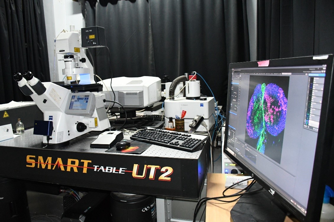
ZEISS LSM 980 AS2 Confocal

equipped

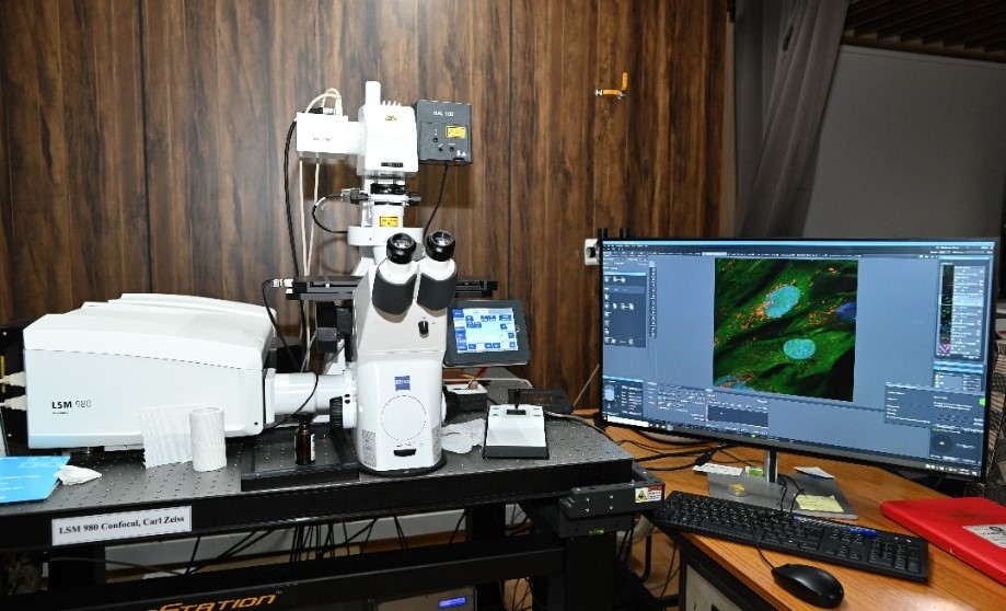
The ZEISS LSM 980 with Airyscan 2 is a next-generation laser scanning confocal microscope designed for high-sensitivity, and super-resolution fluorescence imaging. Featuring the innovative Airyscan 2 detector, the system captures significantly more light and spatial information than conventional confocal microscopes, delivering enhanced resolution, superior signal-to-noise ratio, and improved imaging speed with minimal phototoxicity. Integrated with automated stage control, Definite Focus technology, spectral detection, and live-cell imaging capabilities, the LSM 980 is an ideal platform for advanced biological research.

equipped  equipped  equipped 

Objectives10x/0.3NA, 20x/0.7NA DIC, 40x/1.25NA, 63x/1.4NA Oil, 100x/1.4NA Oil
Lasers405nm, 488nm, 514nm, 561nm, 594nm, 639nm (all solid-state lasers)
DetectorsFour-channel GaAsP PMTs, Two-channel Multi-Alkali PMTs (6 spectral channels), Airyscan 2 detector, and Transmitted Light PMT
SoftwareZEISS ZEN Blue (Version 3.x)
Detection ModesSpectral imaging, Airyscan Super-Resolution, Multiplex imaging, Transmitted light imaging

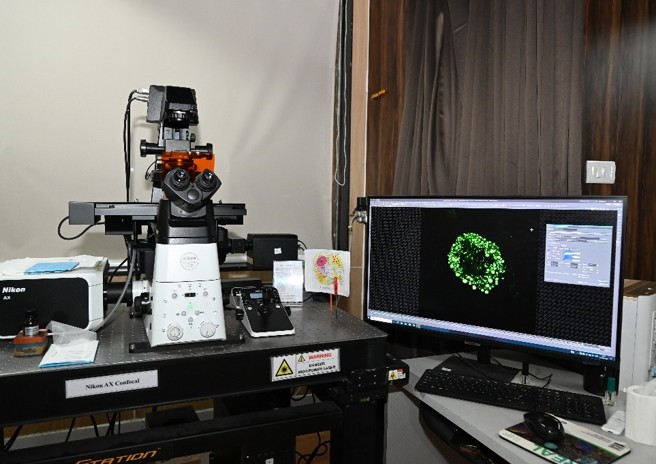
CONFOCAL SYSTEMS Nikon AX Confocal

equipped

The Nikon AX Confocal Microscope is a state-of-the-art laser scanning confocal imaging system engineered for high-speed, high-resolution, and large field-of-view imaging. Built on the Nikon Ti2-Eclipse fully motorized inverted microscope, with 25 mm ultra-large field-of-view galvo scanner, up to 8192 × 8192-pixel image acquisition, imaging speeds up to 10 fps, 360° scan field rotation, Perfect Focus System (PFS) for long-term live-cell imaging, environmental incubator for 35 mm dishes and multiwell plates.

equipped  equipped  equipped 

Objectives10X/0.3NA, 20X/0.7NA DIC, 40X/1.25NA Si (silicon oil),60X/1.4NA Oil, 100X/1.4NA Oil
Lasers405nm, 488nm, 561nm, 640nm (all solid-state lasers)
Detectors4 GASP detectors, one Transmitted Light Detector. Both Channel mode (filter based) and Spectral mode imaging possible
SoftwareNIS Elements AR (Ver.5.4.2) is used both as acquisition and analysis software. Acquisition with XY Overview enables selection of region of interest from a large field of view. Workstation PC includes AI based software innovations like, Auto signal.ai and Denoise.ai and deconvolution to improve resolution.
Imaging ModesBrightfield, Phase contrast, DIC, Fluorescence

LSM780 Confocal

equipped

equipped 

FeaturesAxioObserver Z.1 Inverted microscope attached to confocal scan head, Definite Focus - Autofocus System, Motorized XY Stage, Excite Metal Halide Light Source, RGB filter sets for Epi-fluorescence visualization. The scanning field size can go up to 6144 x 6144 pixels. Rotation of the scan field over 360º is possible. A stage mount incubation chamber (maintained at temperature 37ºC and 5% CO2) can be attached for live cell imaging.
Objectives10x/0.45NA Air (PH1), 20x/0.8NA Air (PH2), 40x/1.3NA Oil, 63x/1.4NA Oil, 100x/1.4NA Oil.
Lasers405nm Diode, Multiline Argon laser 458nm, 488nmn, 514nm lines, DPSS 561nm, HeNe 594m, HeNe 633nm.
DetectorsThe system consists of 3 detectors, 2PMTs and a 34 Channel Spectral Array, all of them can be used for fluorescence and reflection imaging. An additional PMT for transmitted light allows for simultaneous detection of fluorescence and bright field/DIC images.
SoftwareThe system is controlled under LSM Software, ZEN 2.3 (Black) Ver. 14.1. A Workstation PC for analysis with the same software is available, it enables in quantitating length / area / mean fluorescence intensity, colocalization coefficients, assigning different LUTs etc. and exporting of images. Additionally, it helps in FRAP analysis.
Applications of Confocal Microscopy:
  • High-resolution fluorescence imaging of cells and tissues
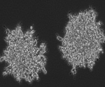
  • Optical sectioning and three-dimensional (3D) reconstruction of cells, tissues, organoids spheroids etc.
  • Live-cell and short and long-term time-lapse imaging to study migration, division, autophagy and dynamics of organelles and proteins.
  • Multi-color fluorescence imaging
  • Protein localization and intracellular trafficking studies
  • Colocalization and interaction analysis of biomolecules (FRET)
  • Fluorescence Recovery After Photobleaching (FRAP) to study mobility and diffusion of proteins.
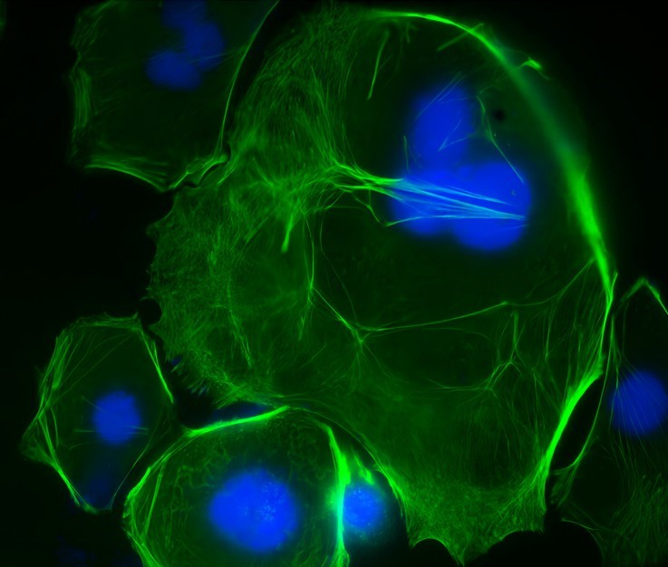
  • Cell morphology and cytoskeletal organization studies
  • Organelle imaging (mitochondria, nucleus, lysosomes, Golgi, ER, etc.)
  • Cancer biology and tumor microenvironment research
  • Drug screening and pharmacological studies
  • Large-area tissue imaging using tile scanning
  • Quantitative fluorescence intensity measurements
  • Biomaterial and scaffold imaging
  • Reflection imaging of non-fluorescent specimens

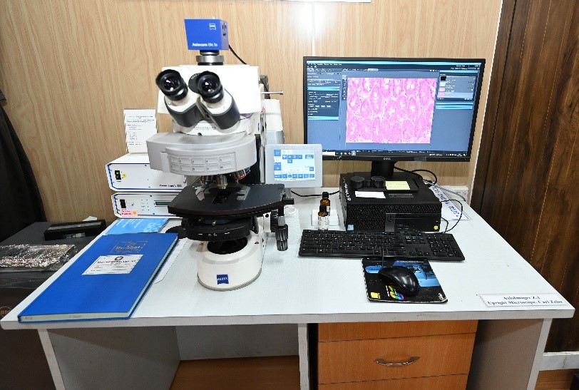
WIDEFIELD SYSTEMS - Axio Imager Z.1 Upright Microscope, Carl Zeiss

equipped


The AxioImager Z.1 is an upright fluorescence microscope used for imaging of cells, tissues, and diverse biological specimens. Partially motorized components, equipped with ZEISS optics - 6 position emission filter wheel and objective turret.

equipped  equipped  equipped 

Objectives5X/0.12NA Air, 10X/0.45NA Air (PH1), 20X/0.8NA Air (PH2), 40X/1.3NA Oil, 40X/0.95NA Air, 63X/1.4NA Oil, 100X/1.4NA Oil.
DetectorsEquipped with a colored camera Axiocam ERc 5s, Carl Zeiss.
SoftwareZen3 PRO (Blue Edition) Ver.3.0
Imaging ModesBrightfield, Phase Contrast and Fluorescence
ApplicationsH&E-stained tissues, Immunohistochemistry (IHC), Fluorescence imaging of cells and tissue sections.

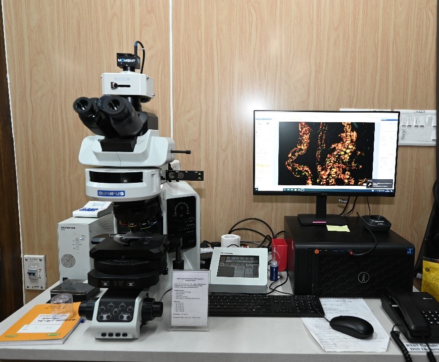
BX63, Upright Microscope, Olympus

equipped

The Olympus BX63 is a partially motorized upright microscope used for imaging of biological specimens - cells, tissues, organism etc. with 8 position emission filter wheel, LED as illumination sources for fluorescence light.

equipped  equipped  equipped 

Objectives10X/0.3NA Air (PH1), 20X/0.7NA Air (PH2), 40X/0.9NA Air, 60X/1.4NA Oil, 100X/1.4NA Oil.
DetectorsMonochrome (Moments, Teledyne Photometrics) Colored camera (DP23, Olympus)
SoftwareCell Sens Dimension Software (Ver. 3.2)
Imaging ModesBrightfield, Fluorescence, Polarization microscopy
ApplicationsH&E stained histopathology tissues, Immunohistochemistry, fluorescently labelled cells and tissue, cells and tissues under polarized light

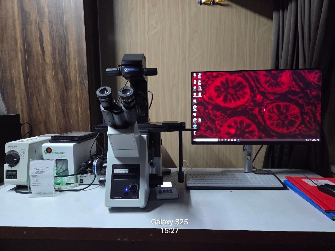
IX73 Inverted Microscope, Olympus

equipped

The Olympus IX73 is a compact inverted fluorescence microscope designed for routine cell culture observation and basic fluorescence imaging. Equipped with phase contrast, fluorescence illumination, and a color digital camera

equipped  equipped 

Objectives10X/0.30 Air, 20X/0.70 Air, 40X/0.65 Air
DetectorsColored camera DP28, Olympus
SoftwareCell Sens Dimension Software (Ver. 3.2)
Imaging ModesBrightfield, Phase contrast, Fluorescence
ApplicationsCell culture observation, Fluorescence microscopy, Phase contrast imaging, Cell morphology analysis, Cell motility and invasion assays Transfection efficiency assessment

Axio Observer Inverted Microscope, Carl Zeiss

equipped

The Carl Zeiss Axio Observer is a fully motorized inverted fluorescence microscope used for advanced live-cell and fluorescence imaging. Featuring an environmental chamber, Colibri LED illumination and Definite focus for z-drift compensation.

equipped  equipped  equipped 

Objectives10X.45 Air (PH1), 20X0.80 Air (PH2), 40X0.95 Air (DIC)
DetectorsHigh-sensitivity AxioCam 802 monochrome camera
SoftwareZEN Pro 2.0 (Blue Edition) software
Imaging ModesBrightfield, Phase Contrast, DIC, Fluorescence, Tile Scan, Time-lapse and Multidimensional imaging
ApplicationsTime-lapse imaging, Tile scan imaging, Transfection efficiency assessment, Multidimensional imaging (XYZCT), Cell migration and proliferation studies

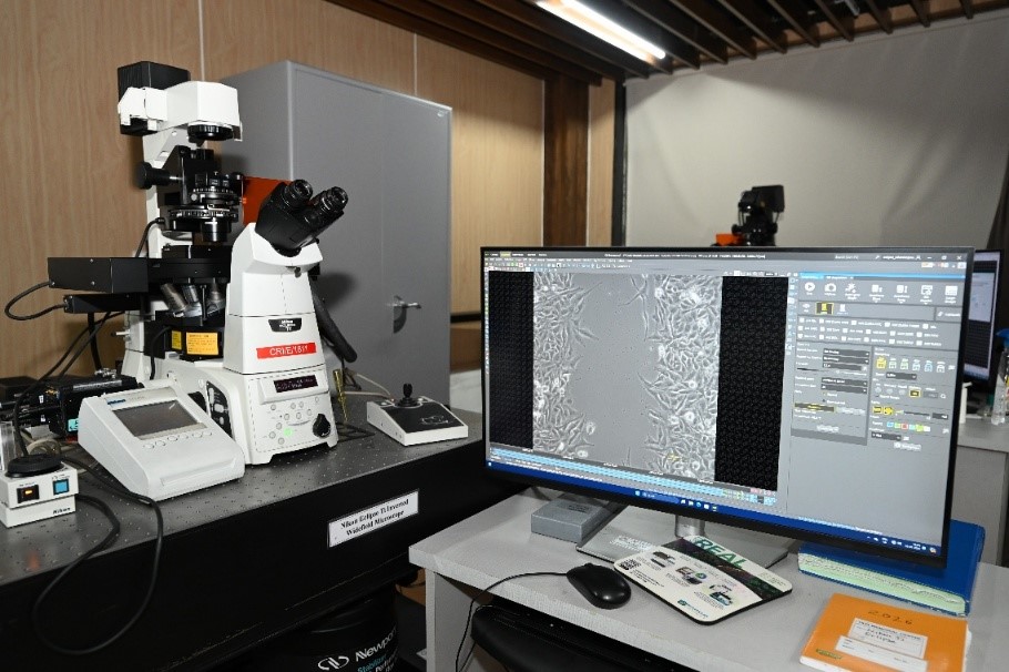
Nikon Eclipse Ti Inverted Microscope

equipped

Nikon Eclipse Ti Inverted Microscope is a Live-cell and fluorescence imaging system. Equipped with an environmental chamber that maintains physiological conditions (37°C and 5% CO₂), the system enables long-term imaging of living cells. It has the Perfect Focus for Z drift compensation.

equipped 

Objectives10X0.30 Air (PH1), 20X0.70 DIC, 20X0.70 Air (PH2)
Detectors2 Monochrome camera - ORCA-Flash4.0 and Andor Clara
SoftwareNIS Elements AR software (Ver.4.2)
Imaging Modes Brightfield, Phase Contrast, DIC, Fluorescence, Tile Scan, Time-lapse
ApplicationsTime-lapse imaging, Wound healing assays, Fluorescence microscopy Cell viability and proliferation , Transfection efficiency studies, Phase Contrast and DIC imaging, Cell motility and invasion assays.

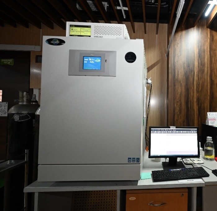
Incucyte

equipped

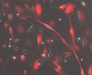
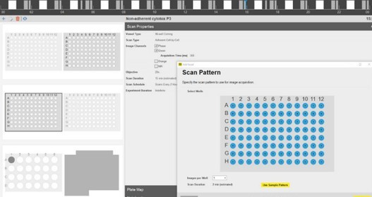
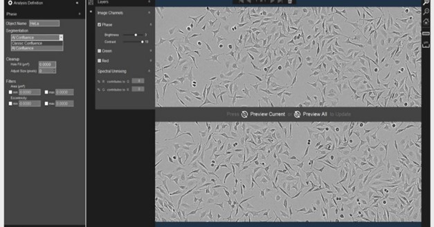
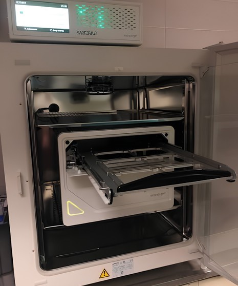
This is a widefield inverted live cell system engineered with long term high throughput imaging in mind. Housed inside a tissue culture incubator, the imaging module maintains the most stable imaging conditions possible (37ºC temperature, humidity and 5% CO2). The system comes with a selection of inserts allowing you to image your cells in a range of vessels (ranging from 35mm dishes to 384 well plates). It automatically captures and analyses images of living cells in real time for days, weeks, or months. It has a optical design wherein the cells remain stationary while optics move. Multiple imaging modes – HD phase plus red and green fluorescence. It can accommodate multiple users at a time with different experiment set ups and configurations


equipped  equipped  equipped  equipped  equipped  equipped 

Objectives4X, 10X, 20X
DetectorsCMOS camera
Imaging ModesPhase contrast, fluorescence (Red and Green only)
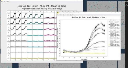
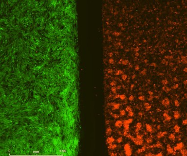
ApplicationsThe acquisition software comes with automated analysis tools to aid with high throughput analysis, such as object counting, scratch wound closure, and confluence measurements. Results are also displayed as plate maps making it easy to see instantly how parameters change between different wells/treatments. You can visualize and validate results by exporting them in the form of images / movies/ data tables. Analysis modules (Essen Incucyte 2020A) available with the system are, Basic Analyzer, Scratch Wound, Chemotaxis, etc.

Dedicated analysis workstations equipped with Imaris, Arivis Pro

Imaris for advanced 3D visualization, segmentation, cell tracking, and quantitative image analysis.

Arivis Pro for AI-assisted segmentation, analysis of large image datasets, multidimensional visualization, and automated workflows.

equipped  equipped 

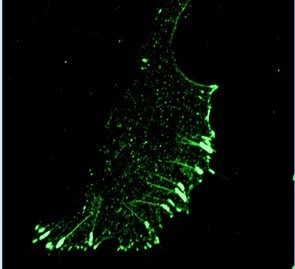
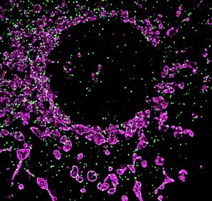
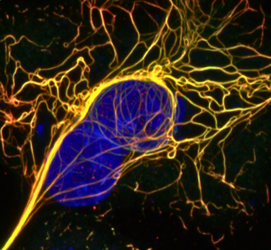
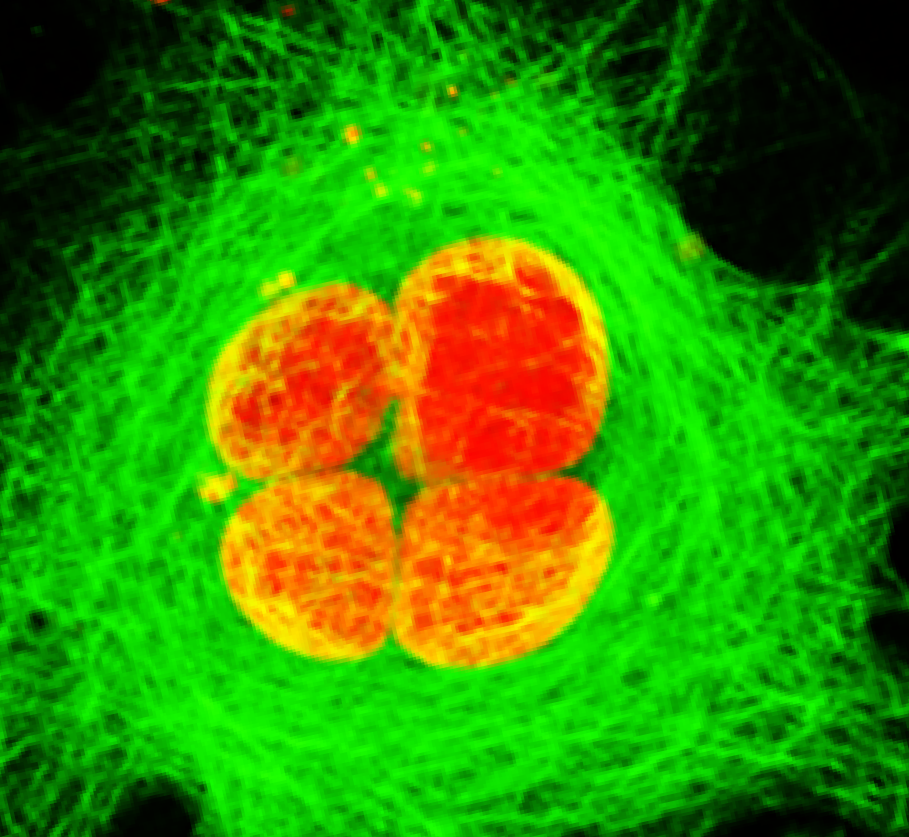
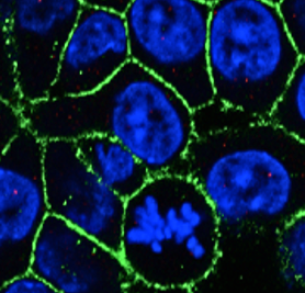
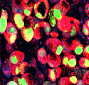
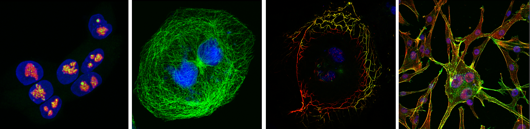
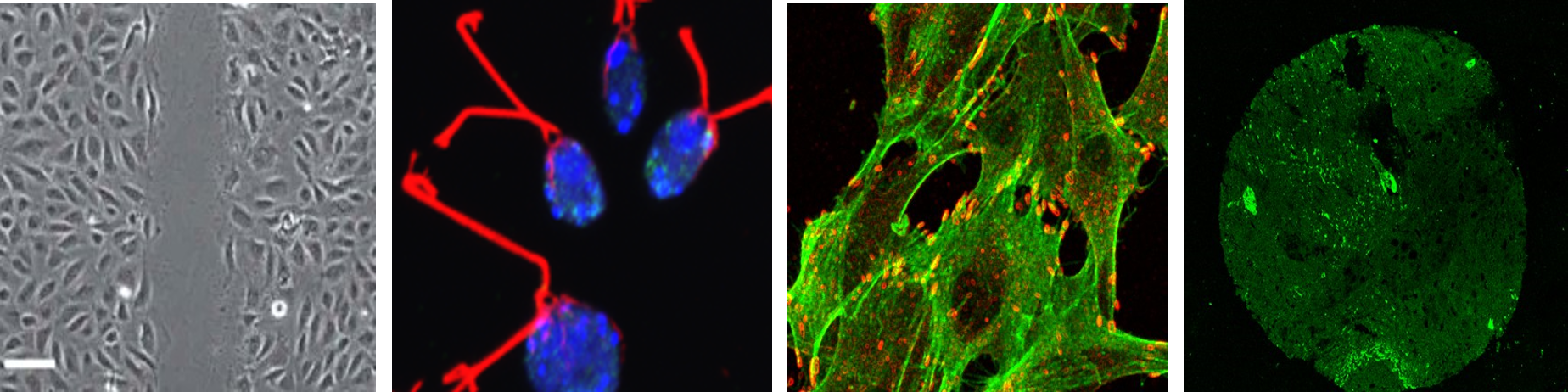
Scenes Beyond the Seen - Understanding Cell Biology through Pixels
CellCell
Services Provided

Advanced Microscopy

Confocal laser scanning microscopy and improved-resolution imaging of subcellular structures with Airy Scan hardware. Multi-channel fluorescence imaging, Spectral imaging and spectral unmixing

Widefield fluorescence microscopy - Brightfield, phase-contrast, and DIC imaging, Tile scan - large-area imaging.
Time-lapse imaging - long-term live-cell imaging under controlled temperature and CO₂ conditions with automated multilocation and multichannel imaging.

Image Processing

Image deconvolution, noise reduction and AI-assisted denoising, spectral unmixing,3D reconstruction and rendering, image stitching, movie generation and image annotation

Quantitative Image Analysis

2D, 3D, and 4D image visualization, cell segmentation and object detection, colocalization analysis, fluorescence intensity measurements, surface, volume, and morphology quantification, AI-assisted image segmentation and analysis.

Functional Imaging

  • Fluorescence Recovery After Photobleaching (FRAP) / FRET (Förster Resonance Energy Transfer)
  • Cell migration and wound-healing assays
  • Chemotaxis and proliferation studies
  • Confluence and viability analysis using Incucyte

User Training and Technical Support

  • Instrument operation training
  • Experimental planning and consultation
  • Assistance in optimizing imaging protocols for diverse biological samples
  • Selection of appropriate imaging techniques
  • Assistance with image acquisition and optimization
  • Image analysis guidance
  • Troubleshooting and technical support

Data Management

  • Secure storage of microscopy data during acquisition
  • Image export in multiple formats
  • Data organization and archiving support
  • Guidance on image processing workflows and data handling
  • Facility Access & User Support

    We are happy to assist you with your microscopy and image analysis requirements. Feel free to contact us by email or phone for any queries related to the facility or our services.

    In-house Certified Users

    Certified in-house users can access the facility independently after regular working hours, as well as on weekends and public holidays, subject to facility guidelines and instrument availability.

    External Users

    Researchers from external institutes are welcome to use the facility. Please email us with a brief description of your project, your imaging requirements, and the microscope(s) you wish to use. Our team will guide you in selecting the most suitable system and assist with scheduling your experiments.

    Media Gallery

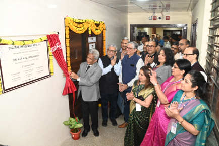
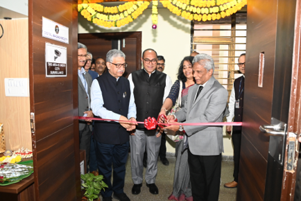
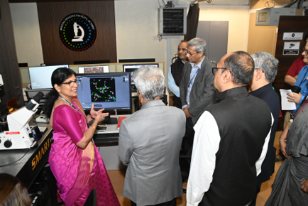
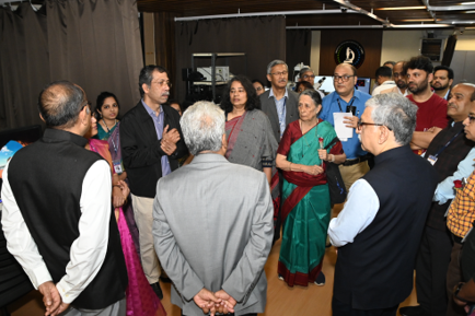
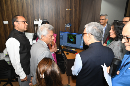
    INAUGURATION OF DIGITAL IMAGING FACILITY (REIMAGINED AND RENOVATED)

    faculty
    faculty
    faculty
    faculty
    faculty

    SCIENCE DAY

    faculty
    faculty
    faculty
    faculty
    faculty
    faculty
    faculty

    Workshop & Visits

    faculty
    faculty
    faculty
    faculty
    Publications

    2026

    2025

    2024

    2023

    2022

    2021

    2020

    2019

    2018

    2017

    2016
    2015

    2014

    2013

    2012

    2011

    2009

    2008

    Acknowledging the Facility

    YOUR RESEARCH OUR SUPPORT

    Your acknowledgement is more than a mention; it is a small gesture of appreciation that encourages and motivates our team to continually improve the quality of our services and support for the researchers.

    If your research has benefited from the support and assistance provided by the Digital Imaging Facility, we kindly request you to acknowledge the Facility in your publications, presentations, theses, and posters.

    Connect with our facility

    Facility Email:adif@actrec.gov.in
    Mrs. Vaishali Kailaje: vaishali.kailaje@actrec.gov.in
    Mrs. Tanuja Durve: tanuja_d29@gmail.com
    Phone no. (+91-22) 2740 5000 or 6873 5000 – Extension 5317/5538

    CIR
    Officer In charge: Mr. Uday Dandekar

    The ‘Instrumentation and Technical Support Department’ serves as a centralised facility for Advanced Scientific Instrumentation and analytical services, designed to support multidisciplinary research in cancer biology. The department provides access to state-of-the-art equipment for characterization, analysis, and testing, enabling students and research scholars to carry out high-quality experimental work efficiently.

    The department aims to promote a culture of shared resources and optimal utilization of equipment by offering sophisticated instruments to in-house departments and external institutions. By integrating cutting-edge technology with expert technical support, the facility enhances research capabilities across diverse labs. In addition to routine analytical services, it facilitates hands-on training to help users gain technical expertise and ensure the optimal use of resources. The department operates on a transparent, user-friendly booking system to maximize accessibility and efficiency.

    All major equipment installed in the department have been covered under an annual maintenance contract in order to provide safe, sustainable, efficient and reliable facilities. The department has a dedicated team of technical experts, who provide necessary technical support to all the equipment installed at other research labs and facilities of the CRI to maintain the equipment in working condition and to reduce downtime of the equipment. Dedicated team of technicians also perform in-house testing, periodic preventive maintenance and calibration, spare part inventory management of the equipment.

    The department also provides technical support in the procurement, installation, and commissioning of the equipment by preparing technical specifications, technical evaluation of the tenders, installation site preparation, warranty claims, asset management, Repair work orders.

    Overall, the ‘Instrumentation and Technical Support Department’ plays a vital role in fostering scientific excellence through the effective utilization of shared research infrastructure.

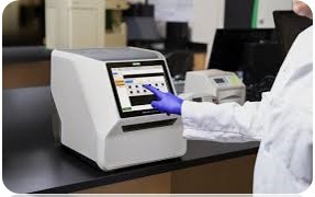
    Microplate Readers

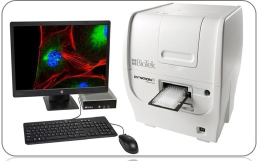
    MULTIMODE MICROPLATE READER - BIOTEK CYTATION 5

    faculty

    A multimode microplate reader is a versatile laboratory instrument that analyzes multiple samples simultaneously in a microplate using multiple detection methods. These complex optical instruments are essential for high throughput screening in the life sciences and pharmaceutical research enabling researchers to quantify biological and chemical reactions and properties in various assays, including drug discovery, cell proliferation, and gene expression

    Technical Features:

    Automation Fully Automatic
    Detection MethodAbsorbance, Luminescence and Fluorescence detection
    Multiple ReadingYes
    Read methodsEndpoint, kinetic, spectral scanning, well area scanning
    Temperature control4-Zone incubation to 65°C with Condensation Control; +0.2°C at 37°C
    Wave length Range230 nm to 999 nm.

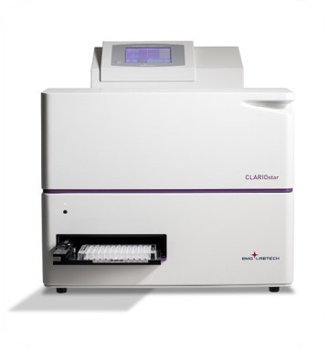
    MULTIMODE MICROPLATE READER - BMG LABTECH CLARIOSTAR PLUS

    faculty

    A wide range of life science applications, including cell based assays, metabolic analysis (oxygen consumption, pH, ROS), enzymatic assays, quantification (RNA, protein), and reporter gene assays. It offers flexibility through high performance monochromator and tunable dichroic mirrors, a dedicated spectrometer for high resolution spectral scanning and atmospheric control to simulate physiological conditions.

    Technical Features:

    Automation Fully Automatic
    Detection MethodAbsorbance, Luminescence and Fluorescence detection
    Multiple ReadingYes
    Read methodsEndpoint, kinetic, spectral scanning, well area scanning
    Temperature control4 Zone incubation to 65°C with Condensation Control; +0.2°C at +37°C
    Wave length Range230 nm to 999 nm.

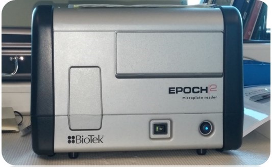
    MICROPLATE READER - BIOTEK EPOCH 2

    faculty

    A microplate reader is a laboratory instrument that quantifies chemical, biological, or physical changes within the wells of a microplate, a multi-well plate used for high-throughput screening. It detects and measures optical signals like absorbance to analyze multiple samples simultaneously, saving time and costs for researchers in the life sciences and pharmaceutical industries.

    Technical Features:

    Automation Automatic
    Multiple ReadingYes
    Read methodsEndpoint, kinetic, spectral scanning, well area scanning
    Wave length Range200 nm to 999 nm.
    Spectrophotometers

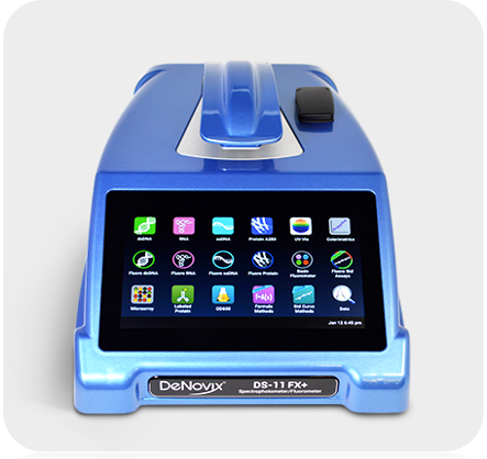
    MICROLITER SPECTROMETER - DENOVIX DS11+

    faculty

    A microliter spectrophotometer is used to quickly measure the concentration and purity of biological samples, such as DNA, RNA, and proteins, by analysing their light absorbance in just 1-2 microliters of liquid. It works by placing a small sample onto a measurement pedestal, where surface tension forms a liquid column between two fibre optics that passes light through it. The instrument then measures the light absorbed at various wavelengths, which indicates the sample's concentration and the presence of contaminants, allowing researchers to assess sample quality for experiments.

    Technical Features :
    Fluorescence UV / Blue, Red, Green
    Absorbance Range190 – 840 nm
    Wavelength accuracy ( Absorbance )0.5 nm
    Micro volume path length0.5 to 0.02 nm
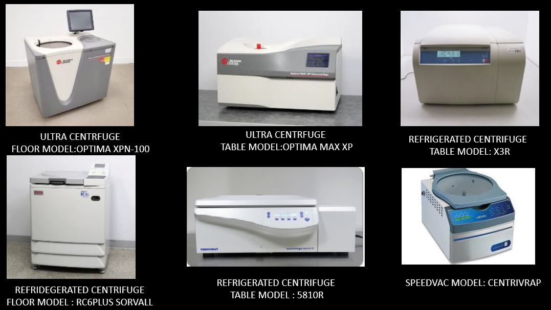
    Centrifuges and Vacuum Concentrators
    faculty
    Gel Documentation Systems

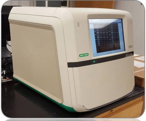
    CHEMIDOC IMAGING SYSTEMS - BIO RAD MP

    faculty

    The ChemiDoc imaging system from Bio Rad is used to image and document gels and western blots. The ChemiDoc imaging system is compatible with a wide range of gel stains, including Ethidium bromide, SYPRO Ruby, Coomassie, and Silver stains. The ChemiDoc Imaging System provides fast, reliable, and sensitive imaging and documentation of gels and Chemiluminescence western blots. This system is compatible with stain free technology.

    Technical Features:

    Smart Tray Technology™ Automatic recognition of application specific tray and adjustment of imaging .
    AutofocusRecalibrated focus for any zoom setting or sample height
    Auto exposureTwo user defined modes (rapid or optimal auto exposure) for all blot and gel imaging applications

    GELDOC IMAGING SYSTEMS - BIO RAD GO IMAGING

    faculty

    The GelDoc Go Imaging System gives you a benchtop imaging solution in a compact and evolved package Acquire high resolution, publication quality images of both nucleic acid and protein gels

    Technical Features:

    Smart Tray Technology™ Automatic recognition of application specific tray and adjustment of imaging .
    AutofocusRecalibrated focus for any zoom setting or sample height
    Auto exposureTwo user defined modes (rapid or optimal auto exposure) for all blot and gel imaging applications
    Shakers and Incubators

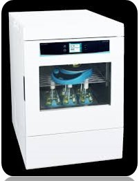
    SHAKER INCUBATOR - LAB COMPANION ISS 3075R

    faculty

    Best space efficient model with double stacking Up to 80°C, 500 rpm enables setting of various experimental conditions. Utilized as an incubator using the shelves Three operating modes Temperature Shaking, Temperature, or Shaking Includes internal power outlet Shaking structure with minimized vibration.

    Technical Features:

    Temperature Range +5°C to +80°C
    Rotation Range (RPM)10 RPM to 180 RPM MAX.

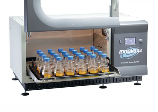
    SHAKER INCUBATOR - INNOVA 44R

    faculty

    A Lab Companion incubator shaker is a laboratory instrument that controls temperature and provides mechanical agitation for incubating samples like microorganisms, cells, or enzymes. These devices are used in fields such as biotechnology and microbiology to facilitate growth and cultivation by providing a controlled environment for heating and shaking at user set speeds and temperatures.

    Technical Features:

    Temperature Range Ambient -10°C to +80°C
    Door openingGlide up
    Speed Range25-400 rpm with 2.5 cm orbit;
    25-300 rpm with 5.1 cm orbit or 2 stacked units;
    25-250 rpm for 2 stacked units
    PCR Machine

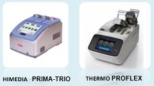
    PCR MACHINE (THERMAL CYCLER) - HIMEDIA AND PROFLEX

    faculty

    A PCR machine amplifies specific DNA or RNA segments from samples, which is essential for applications like detecting genetic diseases, identifying pathogens (e.g. viruses), forensic DNA analysis, paternity testing, and gene cloning. It achieves this by rapidly cycling through controlled temperature changes to replicate target genetic material, a process known as Polymerase Chain Reaction (PCR)

    Technical Features:

    TYPE Multi block Gradient Thermal Cycler
    Block3 x 32 well block
    GUIGraphical touch screen

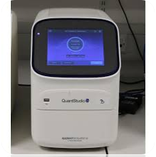
    REAL TIME PCR SYSTEM - QUANT STUDIO 5

    faculty

    The Applied Bio systems Quant Studio 5 Real Time PCR System is designed for Proven OptiPlex technology (six decoupled channels/ 21 filter combinations with white LED) yield optimum results.

    Technical Features:

    Display type touchscreen
    Format384 well plate
    Volume100 µL
    Sonicators and Bioruptors

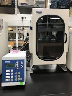
    PROBE SONICATOR - BRANSON 450 DIGITAL

    faculty

    A probe Sonicator is a laboratory instrument that uses high frequency ultrasonic energy to process liquid samples, commonly for disruption, dispersion, and mixing. It works by converting electrical energy into intense vibrations transmitted through a metal probe directly into the liquid, creating cavitation bubbles that cause intense shearing and mixing. This allows for applications like breaking open cells, preparing nanomaterials, and creating emulsions.

    Technical Features:

    Frequency 25KHz
    Pulse ModeAllows for ON and OFF cycles, adjustable from 1 second to 59 seconds, to prevent overheating.
    Power100-1500W

    DIAGENODE SONICATION SYSTEM - BIORUPTOR PICO

    faculty

    A Bioruptor is a laboratory device that uses focused ultrasound and controlled cavitation to create mechanical stress, primarily for shearing biomolecules like DNA and chromatin, but also for disrupting cells, homogenizing tissue, dispersing chemical components, and reducing the size of liposomes.

    Technical Features:

    FREQUENCY 20 KHz-60 KHz
    POWER 1000 watts
    TEMPERATURE +4°C to +20°C
    Educational Video Gallery
    Other Equipment
    faculty

    Dr. Abhijit De
    Facility In Charge: Ms. Snehal Valvi

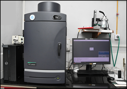
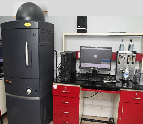
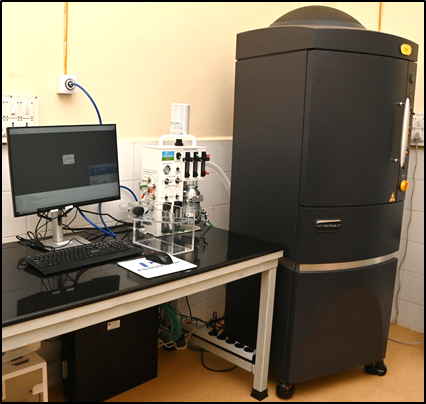
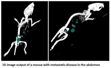
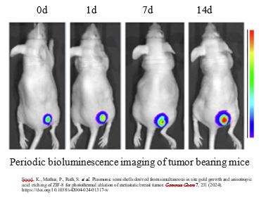
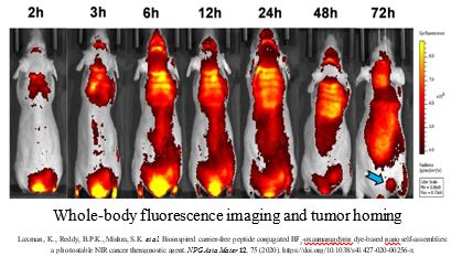
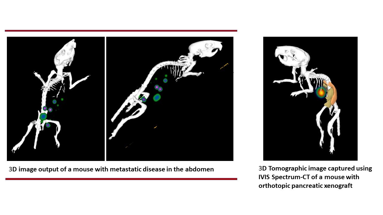
    Considering the growing importance of detecting cancer in a whole-body context of preclinical model organisms that the researchers use in experimental settings, the Molecular Imaging Facility (MIF) was envisioned by Dr. Abhijit De. The MIF facility started its operation in 2013, offering advanced imaging capacity for both live cell population and preclinical in vivo imaging services to researchers. This core facility supports imaging based on three forms of photonic signatures, i.e. Bioluminescence, Cerenkov luminescence and Near InfraRed Fluorescent (NIRF), providing valuable molecular and functional information from whole body context. Current equipment provide ability to scan live cells and small animals to support longitudinal monitoring of disease progression, tracking therapy response and tracking biodistribution of labelled cells and materials. Fast scanning process allows rapid generation of high-quality imaging data for visualization and quantitative analysis. This facility has established track record of supporting imaging work for in-house researchers as well as external research collaborations with institutions and industries.

    Facility Equipment and Imaging Support

    1. IVIS Lumina II (PerkinElmer, USA)

    faculty

    IVIS Lumina II is 2D optical imaging system, both for live cell and live animal imaging in Bioluminescence, Fluorescence and Cherenkov luminescence mode.
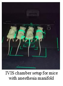
     It allows scanning of upto 3 mice at a time.
     Equipped with four standard fluorescence emission filters GFP, Cy5.5, DsRed and ICG.

    Technical Features:

    Imaging System ComponentsSpecifications
    Camera Sensor Back-thinned, back-illuminated CCD
    CCD Operating Temperature-90 °C
    Min.-Max. Field of View (FOV)A to D (5 cm- 12.5 cm)
    Camera Lens Aperture (F/Stop)f/1 – f/8 (1.5x, 2.5x, 5x, 8.7x magnifications)
    Min. Image Pixel Resolution50 microns
    Excitation Fluorescence Filters
    Emission Fluorescence Filters
    10 (Min. 430 nm, Max. 745 nm with ± 35 nm bandwidth).
    Illumination sourceEpi-illumination
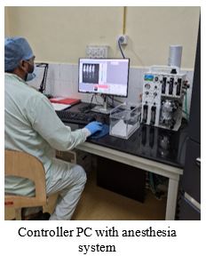
    Imaging Chamber Heated Imaging Stage: 37 °C Integrated gas anaesthesia

    Applications:

    IVIS Lumina II is best suited for routine 2D bioluminescence and fluorescence imaging studies:

    1. Oncology: To monitor tumor growth, metastasis, and anti-cancer therapies in real time using rodent models (Orthotopic, Syngeneic Tumor Models, and Metastasis models).
    2. Gene Expression: To evaluate specific promoter activity and biological process using luciferase reporter genes in vitro.
    3. Infectious Diseases: To check the spread, progression, and clearance of viral or bacterial infections using luminescent pathogens.

    2. IVIS Spectrum (Revvity, USA formerly PerkinElmer, USA)

    faculty

    IVIS spectrum is used for both live cell and live animal imaging in Bioluminescence, Cerenkov luminescence and NIR fluorescence mode.
     Allow scanning of minimum one to maximum five mice per scan in 2D planar mode and 1 mouse for 3D tomographic imaging mode
     Spectral unmixing imaging capacity of IVIS Spectrum provides scope of using multiple probes emitting non-overlapping photonic signatures.
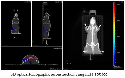
     Uses Structured Light Image to reconstruct the surface tomography of the subject for DLIT and FLIT algorithm.
     Uses transillumination setup to enable effective excitation of deep tissue signal in FLIT mode.

    Technical Features:

    Imaging System ComponentsSpecifications
    Camera SensorBack-thinned, back-illuminated CCD
    CCD Operating Temperature-90 °C
    Min.-Max. Field of View (FOV)A to D (4 cm-22.4 cm)
    Camera Lens Aperture (F/Stop)f/1 – f/8 (1.5x, 2.5x, 5x, 8.7x magnifications)
    Min. Image Pixel Resolution20 microns
    Excitation Fluorescence Filters10 (Min. 430 nm, Max. 745 nm with ± 35 nm bandwidth)
    Emission Fluorescence Filters18 (Min. 500 nm, Max. 840 nm with ± 20 nm bandwidth)
    Illumination sourceEpi-illumination
    Trans-illumination
    Imaging ChamberHeated Imaging Stage: 37 °C
    Integrated gas anaesthesia

    Applications:

    IVIS Spectrum is best suited for routine and advanced multispectral bioluminescence & fluorescence imaging studies:

    1. Pharmacokinetics & Pharmacodynamics: To check the efficacy of various forms of therapy interventions, biodistribution and uptake studies using various radio-isotopes and fluorescent probes in whole animal context.
    2. Protein-protein interaction: Utilizing bioluminescent reporters to study the activation of specific genes and molecular pathways.
    3. Immunology and Stem Cell Research: Observing immune responses, inflammation, and graft-versus-host disease progression longitudinally. Imaging stem cell graft differentiation, testing cell / medical implants in vivo using suitable phantom or model system.

    3. IVIS Spectrum CT (Revvity, USA formerly PerkinElmer, USA)

    faculty

     The IVIS Spectrum-CT is a powerful platform for small animal imaging in pre-clinical settings.
     It provides key optical imaging functionalities: Bioluminescence (BLI), Fluorescence (FLI) and Cerenkov luminescence, for both live cell and live animal imaging.
     High throughput system which allow upto 5 mice in 2D planar scanning mode and upto 2 mouse for 3D scanning mode
     This system provides full 3D tomographic imaging support with co-registered optical signals with precise anatomical (CT) images for volumetric reconstruction of molecular signals.
     Operate in a board spectral range, from visible to near infrared (NIR) light. Spectral unmixing imaging capacity provides scope of using multiple probes emitting non- overlapping photonic signatures.

    Technical Features:

    Imaging System ComponentsSpecifications
    Camera SensorBack-thinned, back-illuminated CCD
    CCD Operating Temperature-90 °C
    Min.-Max. Field of View (FOV)A to D (4 cm-22.4 cm)
    Camera Lens Aperture (F/Stop)f/1 – f/8 (1.5x, 2.5x, 5x, 8.7x magnifications)
    Min. Image Pixel Resolution20 microns
    Excitation Fluorescence Filters10 (Min. 430 nm, Max. 745 nm with ± 35 nm bandwidth)
    Emission Fluorescence Filters18 (Min. 500 nm, Max. 840 nm with ± 20 nm bandwidth)
    Illumination sourceEpi-illumination
    Trans-illumination
    Imaging ChamberHeated Imaging Stage: 37 °C
    Integrated gas anaesthesia
    X-ray Detector Type for Computed TomographyCMOS (Complementary Metal-Oxide-Semiconductor)
    Detector Size3072 x 864 pixels
    Standard Scan DoseMinimum of ~13 mGy

    Applications:

    Common preclinical and biomedical applications include:

    1. Oncology: Enables the longitudinal tracking of tumor growth and metastasis over time. The integrated micro CT then provides 3D anatomical maps (e.g., skeletal structures or lung cavities) to pinpoint the exact location of the tumor.
    2. Angiogenesis Studies: Tracks the formation of new blood vessels feeding tumor growth using near-infrared (NIR) fluorescent targeted or activatable agents.
    3. Bone disease imaging: Combine CT bone anatomy with optical markers of bone remodeling.
    Gallery
    LASACON 2024
    LASACON 2015
    LASACON 2024
    LASACON 2015
    LASACON 2024
    LASACON 2024
    LASACON 2015
    User Guidelines
    • Users are required to submit signed End User Form before starting new experiments. Email the End User at Form to - Email id mifacility[at]actrec[dot]gov[dot]in
    • An online booking software is created to book imaging slots. Users can book slots via URL http://10.100.36.253/FacilityEquipBooking by entering last 6 digits of their CC number and password.
    • Booking slots open a week before; no more than 2 consecutive time slots can be booked by any user.
    • External service users can contact MIF facility directly and discuss requirements.
    Management

    Committee Members -

    Student Members:
    Ms. Diksha Joshi
    Mr. Omkar Dhurat

    Facility In-charge: Ms. Snehal Valvi
    Email: snehal.valvi[at]actrec[dot]gov[dot]in

    Facility Staff: Mr. Amandeep Jast
    Email: jastaman[dot]actrec[at]gmail[dot]com

    Contact Us

    Molecular Imaging Facility
    AH-105 and 107, First Floor, Laboratory Animal Facility, ACTREC, TMC
    Plot No.1 & 2, Sector 22
    Kharghar, Navi Mumbai - 410 210.
    Contact: 022-27405000 / 022-68735000 and ask for extension 5682
    Email: mifacility[at]actrec[dot]gov[dot]in

    Dr. Poonam Gera
    Dr. Poonam Gera

    The ACTREC Biorepository is the custodian of stored biological samples that can be shared, under a tightly regulated and strictly monitored mechanism, with researchers having approved projects that aim to study the biology of cancer, find biomarkers for a more refined molecular classification, or for targeted therapy. In all, tissue samples from 630 cases were accrued in the Biorepository during 2017. As always the majority were head and neck tumors, followed by breast tumors. Other tumor types included neurological, gastrointestinal, genitourinary, gynecological, etc.

    This year, the collection of bio specimens was extended to the Breast OPD wherein core biopsies are now being cryo preserved for future research; this is in addition to collection from the OTs, Frozen room and Surgical Pathology. Cryopreserved tissue samples (470) were provided to eight Principal Investigators with approved projects under various protocols at the Tata Memorial Centre. In the International Cancer Genome Consortium (ICGC) project, gingivo buccal mucosa tumour and blood samples were collected from 30 patients accrued this year, and their extracted genomic DNA samples were sent to NIBMG, Kalyani for whole genome scan and sequence capture-based flow cell sequencing.

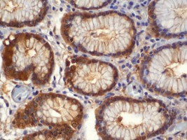
    The Biorepository itself initiated a project on quality monitoring of in-house cryo preserved tissues. The OIC contributed her Pathology expertise involving the evaluation of Hematoxylin & Eosin as well as immunohistochemistry slides to eight ACTREC projects.

    Dr. Ashok Varma
    Dr. Ashok Varma

    The Bioinformatics facility of ACTREC provides infrastructural and technical support to scientists, clinicians and research scholars of the Centre to fulfil the bioinformatics requirements of their on-going research projects. Scientists of the Centre also use the facility’s infrastructure to explore microarray, next generation sequence data analysis, database development, molecular modelling and data mining for their on-going projects.

    This facility receives strong funding support from DBT and is established as a BTIS-net centre of this region. It is well equipped with one nVIDIA Tesla GPU workstation, 5 workstations, 1 webserver and seven PCs. The facility also focuses on database development such as Histome: the human infobase; this database is presently being updated. In the domain of gene expression studies, projects related to TGCA database mining and analysis are under progress. In silico molecular modelling, dynamics and protein-protein interactions have been performed using BARC’s supercomputing facility.

    The facility hosted a national 28th BTISnet Coordinators Meeting on 3rd and 4th February 2017. The facility also organized its annual 2-day Workshop on ‘Basics of Bioinformatics’ targeting college teachers and research scholars of institutions in the Mumbai and neighboring areas on 2nd and 3rd March 2017. The facility staff also provided training to six trainees during the year, three for Bachelor’s/ Master’s dissertation and three for experience.

    Dr K Nirmal Kumar
    Officer In Charge: Dr K Nirmal Kumar

    The Anti-Cancer Drug Screening Facility (ACDSF) at ACTREC supports the efforts of anti-cancer drug development in India, with in vitro and in vivo anti-cancer drug screening assays that have been developed in-house. ACDSF has 53 human tumor cell lines, 10 murine tumor models and 38 xenograft models for carrying out drug screening. During 2017, 1345 compounds were received from 161 clients including eight corporate R&D organizations from 13 states across India. In all, 1287 compounds were tested for their in vitro activity and 58 compounds were examined for MTD (n=14) and in vivo efficacy assays (n=44).

    Two new xenografts namely KG-1 (leukemia) and HCC1954 (breast cancer) were developed during the report year. The facility has successfully completed XII-plan CSIR funded project ‘Affordable cancer therapeutics’ in collaboration with IICT, Hyderabad (2012-17). Of the second set of 300 compounds, 11 compounds were found to be active against four cancer cell lines (Hep-G2, HT-29, SCC-29B and PLC-Prf-5).

    Three of these compounds that were most active against oral cancer spheroids (CSC) were further tested for their in vivo efficacy against oral cancer xenograft AW13516. Only one of these (AKL-JA) was found to be strongly active against the AW13516 xenograft. Corroborative evidence was obtained through PET-CT imaging of tumor bearing animals and histopathology of tumor sections.
    Contact Us

    For sample submission and enquiry: Please write to the facility in charge for further information on service provided, sample submission and requisition form will be provided by the facility. Users are requested to discuss over email/telephone regarding the study and other information.

    Contact:
    Facility in charge: Dr K Nirmal Kumar,
    Scientific Officer ‘D’
    Anti-Cancer Drug Screening,
    Room No. KS-324,
    Advanced Centre for Treatment, Research & Education in Cancer (ACTREC),
    Tata Memorial Centre,
    Kharghar, Navi Mumbai - 410 210.
    Email: acdsf@actrec.gov.in
    Tel: 022-27405000 / 022-68735000
    Ext: 5431

    Back to Top