

Molecular Imaging
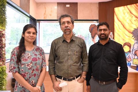
Considering the growing importance of detecting cancer in a whole-body context of preclinical model organisms that the researchers use in experimental settings, the Molecular Imaging Facility (MIF) was envisioned by Dr. Abhijit De. The MIF facility started its operation in 2013, offering advanced imaging capacity for both live cell population and preclinical in vivo imaging services to researchers. This core facility supports imaging based on three forms of photonic signatures, i.e. Bioluminescence, Cerenkov luminescence and Near InfraRed Fluorescent (NIRF), providing valuable molecular and functional information from whole body context. Current equipment provide ability to scan live cells and small animals to support longitudinal monitoring of disease progression, tracking therapy response and tracking biodistribution of labelled cells and materials. Fast scanning process allows rapid generation of high-quality imaging data for visualization and quantitative analysis. This facility has established track record of supporting imaging work for in-house researchers as well as external research collaborations with institutions and industries.
1. IVIS Lumina II (PerkinElmer, USA)
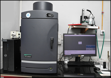
IVIS Lumina II is 2D optical imaging system, both for
live cell and live animal imaging in Bioluminescence,
Fluorescence and Cherenkov luminescence mode.
It allows scanning of upto 3 mice at a time.
Equipped with four standard fluorescence emission filters GFP, Cy5.5, DsRed and ICG.
Technical Features:
| Imaging System Components | Specifications |
|---|---|
| Camera Sensor | Back-thinned, back-illuminated CCD |
| CCD Operating Temperature | -90 °C |
| Min.-Max. Field of View (FOV) | A to D (5 cm- 12.5 cm) |
| Camera Lens Aperture (F/Stop) | f/1 – f/8 (1.5x, 2.5x, 5x, 8.7x magnifications) |
| Min. Image Pixel Resolution | 50 microns |
| Excitation Fluorescence Filters Emission Fluorescence Filters | 10 (Min. 430 nm, Max. 745 nm with ± 35 nm bandwidth). |
| Illumination source | Epi-illumination |
| Imaging Chamber | Heated Imaging Stage: 37 °C Integrated gas anaesthesia |
Applications:
IVIS Lumina II is best suited for routine 2D bioluminescence and fluorescence imaging studies:
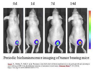
- Oncology: To monitor tumor growth, metastasis, and anti-cancer therapies in real time using rodent models (Orthotopic, Syngeneic Tumor Models, and Metastasis models).
- Gene Expression: To evaluate specific promoter activity and biological process using luciferase reporter genes in vitro.
- Infectious Diseases: To check the spread, progression, and clearance of viral or bacterial infections using luminescent pathogens.
2. IVIS Spectrum (Revvity, USA formerly PerkinElmer, USA)
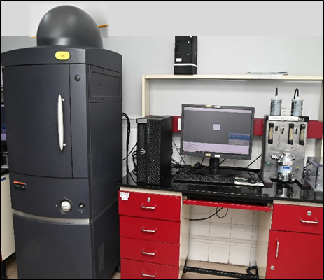
IVIS spectrum is used for both live cell and live
animal imaging in Bioluminescence, Cerenkov
luminescence and NIR fluorescence mode.
Allow scanning of minimum one to maximum five
mice per scan in 2D planar mode and 1 mouse for
3D tomographic imaging mode
Spectral unmixing imaging capacity of IVIS
Spectrum provides scope of using multiple probes
emitting non-overlapping photonic signatures.
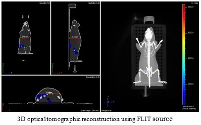
Uses Structured Light Image to reconstruct the
surface tomography of the subject for DLIT and
FLIT algorithm.
Uses transillumination setup to enable effective
excitation of deep tissue signal in FLIT mode.
Technical Features:
| Imaging System Components | Specifications |
|---|---|
| Camera Sensor | Back-thinned, back-illuminated CCD |
| CCD Operating Temperature | -90 °C |
| Min.-Max. Field of View (FOV) | A to D (4 cm-22.4 cm) |
| Camera Lens Aperture (F/Stop) | f/1 – f/8 (1.5x, 2.5x, 5x, 8.7x magnifications) |
| Min. Image Pixel Resolution | 20 microns |
| Excitation Fluorescence Filters | 10 (Min. 430 nm, Max. 745 nm with ± 35 nm bandwidth) |
| Emission Fluorescence Filters | 18 (Min. 500 nm, Max. 840 nm with ± 20 nm bandwidth) |
| Illumination source | Epi-illumination Trans-illumination |
| Imaging Chamber | Heated Imaging Stage: 37 °C Integrated gas anaesthesia |
Applications:
IVIS Spectrum is best suited for routine and advanced multispectral bioluminescence & fluorescence imaging studies:
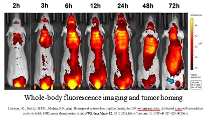
- Pharmacokinetics & Pharmacodynamics: To check the efficacy of various forms of therapy interventions, biodistribution and uptake studies using various radio-isotopes and fluorescent probes in whole animal context.
- Protein-protein interaction: Utilizing bioluminescent reporters to study the activation of specific genes and molecular pathways.
- Immunology and Stem Cell Research: Observing immune responses, inflammation, and graft-versus-host disease progression longitudinally. Imaging stem cell graft differentiation, testing cell / medical implants in vivo using suitable phantom or model system.
3. IVIS Spectrum CT (Revvity, USA formerly PerkinElmer, USA)
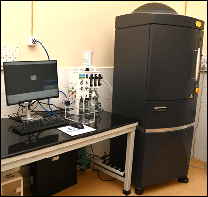
The IVIS Spectrum-CT is a powerful platform for small
animal imaging in pre-clinical settings.
It provides key optical imaging functionalities:
Bioluminescence (BLI), Fluorescence (FLI) and
Cerenkov luminescence, for both live cell and live
animal imaging.
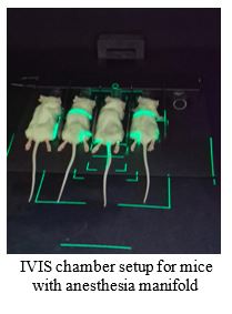
High throughput system which allow upto 5 mice in
2D planar scanning mode and upto 2 mouse for 3D
scanning mode
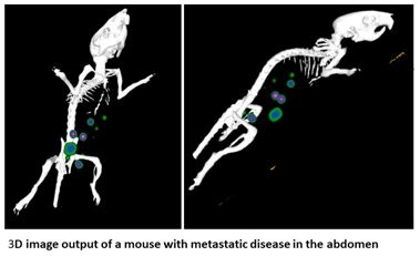
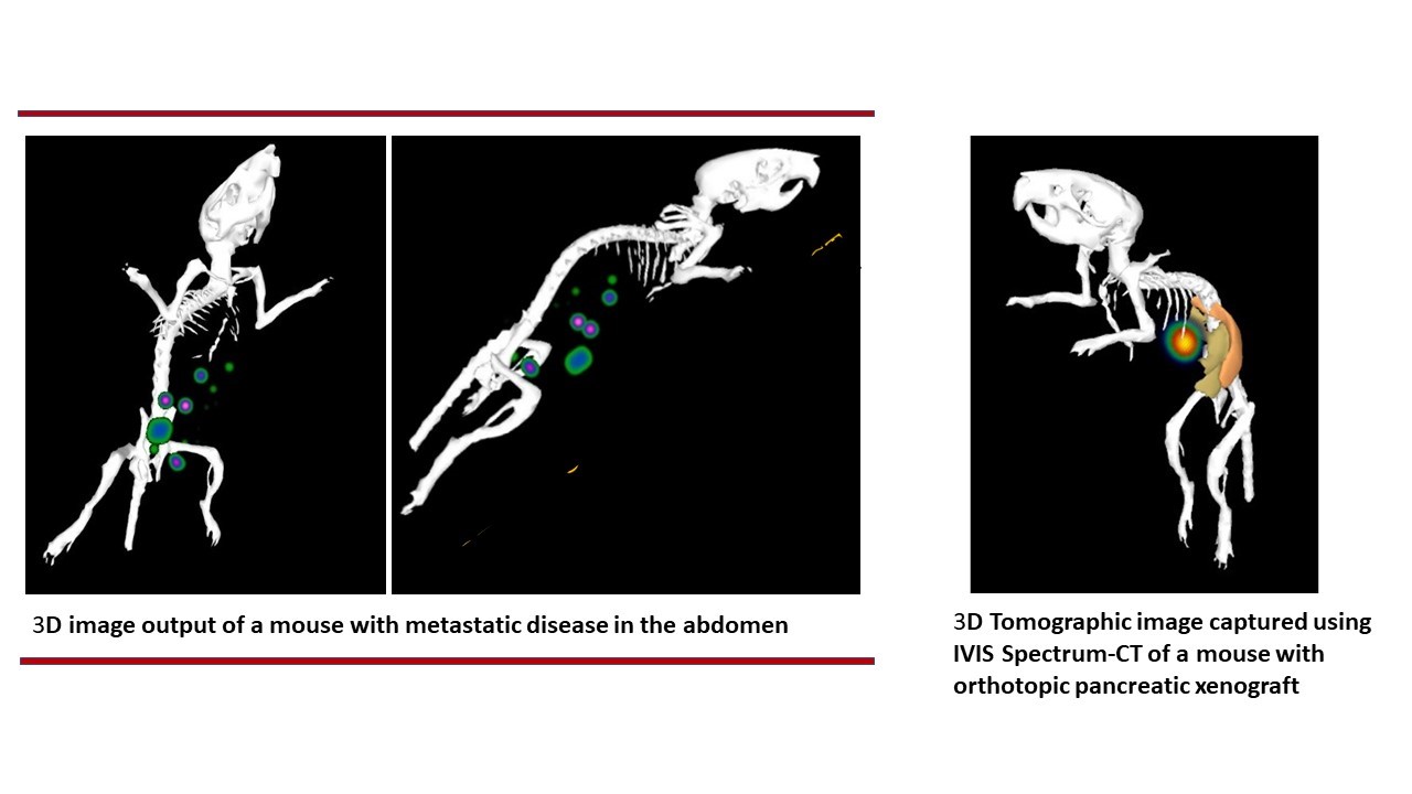
This system provides full 3D tomographic imaging
support with co-registered optical signals with precise
anatomical (CT) images for volumetric reconstruction of
molecular signals.
Operate in a board spectral range, from visible to near
infrared (NIR) light. Spectral unmixing imaging capacity
provides scope of using multiple probes emitting non-
overlapping photonic signatures.
Technical Features:
| Imaging System Components | Specifications |
|---|---|
| Camera Sensor | Back-thinned, back-illuminated CCD |
| CCD Operating Temperature | -90 °C |
| Min.-Max. Field of View (FOV) | A to D (4 cm-22.4 cm) |
| Camera Lens Aperture (F/Stop) | f/1 – f/8 (1.5x, 2.5x, 5x, 8.7x magnifications) |
| Min. Image Pixel Resolution | 20 microns |
| Excitation Fluorescence Filters | 10 (Min. 430 nm, Max. 745 nm with ± 35 nm bandwidth) |
| Emission Fluorescence Filters | 18 (Min. 500 nm, Max. 840 nm with ± 20 nm bandwidth) |
| Illumination source | Epi-illumination Trans-illumination |
| Imaging Chamber | Heated Imaging Stage: 37 °C Integrated gas anaesthesia |
| X-ray Detector Type for Computed Tomography | CMOS (Complementary Metal-Oxide-Semiconductor) |
| Detector Size | 3072 x 864 pixels |
| Standard Scan Dose | Minimum of ~13 mGy |
Applications:
Common preclinical and biomedical applications include:
- Oncology: Enables the longitudinal tracking of tumor growth and metastasis over time. The integrated micro CT then provides 3D anatomical maps (e.g., skeletal structures or lung cavities) to pinpoint the exact location of the tumor.
- Angiogenesis Studies: Tracks the formation of new blood vessels feeding tumor growth using near-infrared (NIR) fluorescent targeted or activatable agents.
- Bone disease imaging: Combine CT bone anatomy with optical markers of bone remodeling.
 |  | |
 |  | |
 | ||
 |  |
- Users are required to submit signed End User Form before starting new experiments. Email the End User at Form to - Email id mifacility[at]actrec[dot]gov[dot]in
- An online booking software is created to book imaging slots. Users can book slots via URL http://10.100.36.253/FacilityEquipBooking by entering last 6 digits of their CC number and password.
- Booking slots open a week before; no more than 2 consecutive time slots can be booked by any user.
- External service users can contact MIF facility directly and discuss requirements.
Committee Members -
- Dr. Abhijit De
Principal Investigator and Scientific Officer ‘H’
Email: ade[at]actrec[dot]gov[dot]in - Dr. Syed Hasan
Principal Investigator and Scientific Officer ‘F’
Email: shasan[at]actrec[dot]gov[dot]in
Student Members:
Ms. Diksha Joshi
Mr. Omkar Dhurat
Facility In-charge: Ms. Snehal Valvi
Email: snehal.valvi[at]actrec[dot]gov[dot]in
Facility Staff: Mr. Amandeep Jast
Email: jastaman[dot]actrec[at]gmail[dot]com
Molecular Imaging Facility
AH-105 and 107, First Floor, Laboratory Animal Facility, ACTREC, TMC
Plot No.1 & 2, Sector 22
Kharghar, Navi Mumbai - 410 210.
Contact: 022-27405000 / 022-68735000 and ask for extension 5682
Email: mifacility[at]actrec[dot]gov[dot]in