
EGFR mutation frequency in Indian lung cancer patients
Clinical collaborator: Dr. Kumar Prabhash (TMH)Here, as a first study from India, we report 23% incidence of EGFR mutations in exon 18-21 by TaqMan probe-based assays in 907 NSCLC patients. Interestingly, an intermediate EGFR mutation rate of 23% among modern-day Indians compared to 10-15% in Caucasians and 27-62% among East Asians is reminiscent of an admixture of genetic influence from Middle Easterners, Central Asians, and Europeans due to different degree of migration wave and amalgamation of major ancestral populations over past thousand of years at different points of time. Furthermore, unlike in developed countries no significant difference were observed between never-smoker adenocarcinoma females and males indicating lack of gender bias among never-smoker patients, unlike in developed countries. Taken together, this study may be critical to plug the crucial deficit in designing of genome wide association studies to determine haplotype that may confer differential susceptibility to somatic mutations in EGFR in NSCLC, as reported for somatic mutations in JAK2 in human myeloproliferative neoplasms.
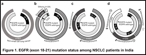
EGFR (exon 18-21) mutation status among NSCLC patients in India
(A) EGFR mutation status across all NSCLC samples is shown. Black segment in the inner circle indicate patients harboring EGFR mutation, the segment in white indicates patients with wild type EGFR. The segment in grey (outer circle) indicates total number of NSCLC patients.
(B) EGFR mutations across different histological subtypes. Grey segment in the outer circle indicate total number of lung adenocarcinoma patients. The patients with squamous histology are represented in dark grey. The segment in white represents lung cancer patients with tumors histology other than adeno or squamous carcinoma. Similar to (A), the black segment in the inner circle indicate patients harboring EGFR mutation, the segment in white indicates patients with wild type EGFR.
(C) EGFR mutation variation among male and female lung adenocarcinoma patients. The light grey slice represents male patients; female lung adenocarcinoma patients represented in dark grey in outer circle. Similar to (A), the black segment in the inner circle indicate patients harboring EGFR mutation, the segment in white indicates patients with wild type EGFR among male and female lung adenocarcinoma patients.
(D) EGFR mutation variation with respect to gender specific smoking habits among all lung adenocarcinoma patients. Light grey segment in the outer circle indicate male adenocarcinoma lung cancer patients with smoking history. The non smoker male and female lung adenocarcinoma patients are represented in dark grey and white, respectively. The segment in white represents lung